Article

Immediate Effect of a Kinesiotape Bandage on Knee Mechanics during Functional Tests in Female Rugby and Football Athletes: A Pilot Study

by
Amandine Fevre
,
Juliette Moriceau
,
Jaime Almazán-Polo
and
Guillermo García-Pérez-de-Sevilla
*
Faculty of Sport Sciences, Universidad Europea de Madrid, Calle Tajo s/n, Villaviciosa de Odón, 28670 Madrid, Spain
*
Author to whom correspondence should be addressed.
Appl. Sci. 2024, 14(7), 2839; https://doi.org/10.3390/app14072839
Submission received: 22 February 2024 / Revised: 22 March 2024 / Accepted: 26 March 2024 / Published: 28 March 2024
(This article belongs to the Special Issue Exercise, Fitness, Human Performance and Health)

Abstract

:
Background: Some bandages with Kinesiotape have demonstrated some potential as means of prevention for Anterior Cruciate Ligament (ACL) injury in men. Objective: The main objective of this pilot study was to observe if a Kinesiotape bandage at the knee could potentially have an immediate preventive effect against ACL injuries by improving proprioception and balance and reducing knee valgus and anterior translation of the tibia during certain functional tests in female athletes. Materials and Methods: A cross-over clinical trial including 10 female athletes (football and rugby) was conducted, where the two lower limbs of the 10 participants (n = 20) were randomly assigned to the intervention group (IG) or to the control group (CG). A Kinesiotape bandage was placed on one knee with a tension of 75% (IG, n = 10) and another bandage on the other knee with a tension of 10% (CG, n = 10), as a placebo. Ultrasound assessment and functional tests were performed before (T1) and after (T2) bandaging according to group assignment. A videographic analysis was carried out with the ImageJ Software version 2.0. Results: A statistically significant reduction in knee valgus was observed in the Lateral Step Down test in the IG, compared to the CG (p < 0.05; η2p = 0.26) (IG-T1: 151.40 ± 11.04°; IG-T2: 157.10 ± 10.18°; versus CG-T1: 156.96 ± 5.44°; CG-T2: 158.68 ± 6.12°). In the other tests, no significant differences were found in terms of time × group interaction. Conclusions: A Kinesiotape bandage was able to reduce knee valgus in a functional test compared to a placebo bandage but was not more effective at reducing the anterior translation of the tibia or improving balance or proprioception. Based on these results, it could have a modest preventive effect against an ACL injury in female athletes.

1. Introduction

The anterior cruciate ligament (ACL) is the main static stabilizer of anterior translation of the tibia and represents up to 86% of the total force resisting anterior displacement. This ligament has a key proprioceptive function because of its numerous mechanoreceptors [1].
The risk of ACL injury is multifactorial, involving biomechanical, anatomical, hormonal, and neuromuscular factors. Of all cases, 70–80% occur without contact. One of the most described mechanisms in women involves landing with the hip and knee extended, knee valgus, tibial internal rotation, and foot pronation [2,3]. ACL injury is one of the most common knee injuries in athletes. Each year in the United States, there are an estimated 250,000 ACL tears [4].
Currently, some studies show that women are more susceptible to ACL injury than men [5,6]. In fact, the number of non-contact injuries is two to nine times higher in women than in men [7,8]. These injuries are more common in young female athletes who play team sports [7] which involve jumping, turning, and changing directions. By sport, the proportion of non-contact ACL injuries to total ACL injuries was 66% in soccer and 54% in rugby [8].
The impact of these injuries on athletes’ sports participation and general well-being can be significant, both in the short and long term. Only 65% of injured soccer players can compete at the same level 3 years after the injury [9,10]. In the long term, ACL injury is associated with potential complications, including chronic knee instability, meniscus tears, cartilage damage, and a seven-fold increased risk of end-stage osteoarthritis (OA) leading to knee replacement [8]. In addition, after ACL reconstruction, less than 50% of patients will return to sports within one year, less than 65% within two years, 24% will change sports, and 11% will drop out [11,12].
Several programs based on the neuromuscular system, such as FIFA11+ [13], achieved a 50% reduction in the risk of ACL injuries in men and 67% in women [14], but a consensus has not yet been established. Knowing that these prevention programs are carried out in a period of 6 to 8 weeks, it would be novel to find a form of ACL injury prevention with an immediate effect.
Among the most-used bandages, Kinesiotape (KT) is proposed as a beneficial therapeutic tool in the prevention and treatment of sports injuries [15,16], helping to reduce pain, increase ROM, and improve proprioception as well as muscle activity [17]. Through tactile input, KT stimulates cutaneous mechanoreceptors and motoneurons [18,19], which can explain why KT has shown positive results in improving function, stability, proprioception, and force production of the muscle in randomized controlled trials [20].
In individuals with a recent ACL rupture, KT application at the knee has been shown to improve proprioception, strength, and static balance [21,22,23]. However, in healthy young athletes, there are inconclusive results about the effectiveness of KT at the knee. In some randomized controlled trials, KT is not more effective than sham KT in improving static and dynamic stability [24]. However, during landing tasks, some studies found that KT at the knee reduces the dynamic knee valgus, which could reduce the risk of an ACL injury, considering that a pronounced knee valgus is a relevant factor [20,25]. In this line, a recent study applied a KT bandage to the knee with a tension of 75%, achieving a significant reduction in the knee valgus during the Jump Landing Test in men [26]. To date, there is inconclusive evidence that KT improves neuromuscular control at the knee in symptomatic populations [27], and it would be interesting to analyze the effectiveness of KT in reducing knee valgus in several closed kinetic chain functional tests focusing on women, since they have a greater risk of suffering from ACL injuries.
Therefore, the main objective of this study was to determine whether a KT bandage at the knee improves balance and proprioception and decreases anterior translation of the tibia and knee valgus in functional tests in female athletes. Our hypothesis suggests that significant differences will be observed in tibiofemoral displacement, as well as in evaluating knee valgus.

2. Materials and Methods

2.1. Study Design, Settings, and Ethical Aspects

A pilot study was conducted using a longitudinal, prospective, randomized, cross-over clinical trial design, following the TREND (Transparent Reporting of Evaluations with Nonrandomized Designs) guidelines [28]. The study was carried out in the physical performance laboratory of the Faculty of Physical Activity, Sports Sciences, and Physiotherapy of the European University of Madrid. The Clinical Research Ethics Committee of the European University of Madrid (SPAIN) authorized the study on 13 February 2023 (CIPI/23.052). Prior to commencing the study, all participants provided informed consent. Moreover, throughout the entire study, strict adherence to the principles outlined in the Declaration of Helsinki was maintained.

2.2. Sample Size Calculation

The sample size was calculated with the G*Power 3.1.9.7 software using an alpha error of 0.05 and a beta of 0.80, considering the total antero-posterior displacement of the tibia as the primary variable. The total needed sample calculated was n = 50 (25 players).

2.3. Participants and Recruitment

To participate in this study, the participants needed to be female amateur rugby or football athletes aged 18–35 years. The exclusion criteria were (a) chronic pathology that prevents physical exercise; (b) pathologies in the lower limbs in the last six months or a history of ACL injury, (c) and skin pathologies that contraindicate the use of bandages (e.g., eczema or psoriasis) (Table 1).
Height (cm) and weight (kg) were measured, and then the body mass index (BMI, kg/m2) was calculated. In addition, the International Physical Activity Questionnaire (IPAQ) was used to evaluate the physical activity levels of the participants (MET’S/min/week) [29]. Finally, the researchers recorded the dominant leg and measured the height of each leg with a tape measure.
Of the 15 women initially recruited, 5 (33%) did not meet the inclusion criteria, so a total of 10 female football or rugby athletes (n = 5 football players; n = 5 rugby players) aged 21.90 ± 3.48 years, with a mean BMI of 21.09 ± 2.55 kg/m2 and 90% right lower limb dominance, were recruited in this pilot study.
According to the IPAQ questionnaire, 50% of the participants had a high level of physical activity and the other half had a moderate level, with a mean of 2861.10 ± 949.62 MET/min/week (Table 1).

2.4. Randomization

The two lower limbs of the 10 participants (n = 20) were randomly assigned to the intervention group (IG) or to the control group (CG). The randomization of the lower limbs to the IG or the CG was performed using the randomization function of Microsoft Excel version 18.0 to prevent the lower limb dominance of each participant from affecting the results.
A Kinesiotape bandage was placed on one knee with a tension of 75% (IG, n = 10) and another bandage on the other knee with a tension of 10% (CG, n = 10), as a placebo. To avoid the risk of assignment, the investigators applied the bandage with 10% or 75% tension randomly to the left or right lower limb.

2.5. Variables

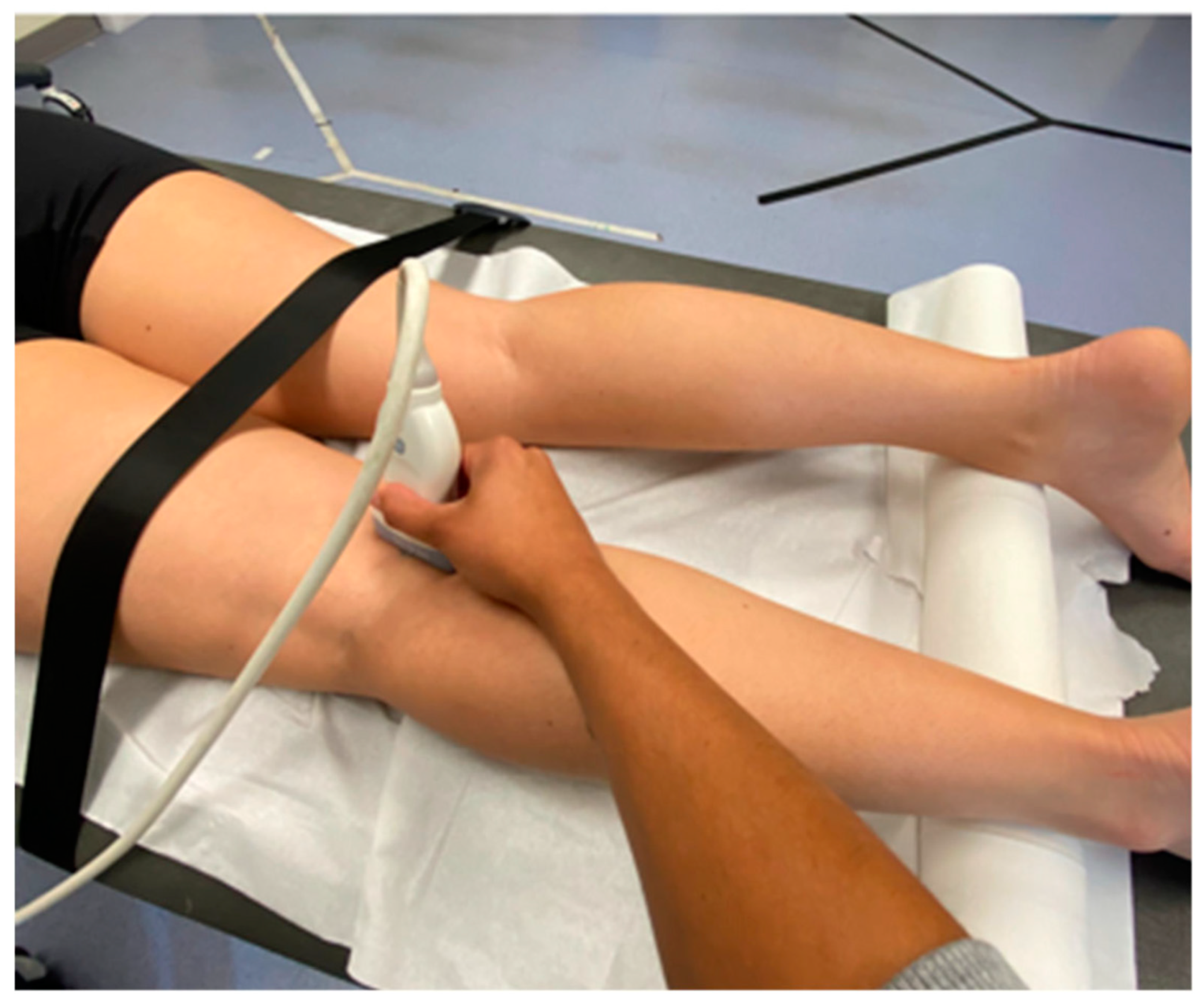
2.5.1. Anteroposterior Displacement of the Tibia Measured with Ultrasound

To perform the ultrasonographic assessment at the knee, an ultrasound machine (Logiq S7 Expert, GE Healthcare, Chicago, IL, USA) was used, equipped with a linear probe (50 mm field of vision) with a frequency range of 4 to 15 MHz. A preset frequency of 8 MHz, 61 gain points, 69 points of dynamic range, and 1 focus located at a depth of 5 cm were established to evaluate the knee morphology. The participants were positioned in the prone position, with a twist under the legs to maintain the knee in 20° of flexion and the femur supported by a McKenzie tape to assess movements of the tibia only. This evaluation technique to was used by Stoianov et al. to observe the accuracy and reliability of dynamic ultrasound assessment of anterior tibial translation (Figure 1) [30].
Measurements were performed on both knees, on the posteromedial side. For the interpretation of the ultrasound data, the ultrasound machine was used directly as a reference. The distance between the tangent line to the medial femoral condyle and the tangent line to the posterior aspect of the tibia was measured in the static position (D1) and after applying manual pressure at the level of the proximal posterior aspect of the tibia (D2) (Figure 2). For each knee, the difference between the two distances (D2 − D1) was calculated. Participants were instructed to keep the leg as relaxed as possible during the ultrasound evaluation. Likewise, the position of the probe was stabilized by firmly maintaining the soundproofing angle as well as maintaining the least possible pressure on the posterior aspect of the knee.
Immediately after, a similar pressure was applied on the proximal anterior aspect of the tibia (D3) to calculate the total displacement of the tibia. Due to this pressure, the full range of anteroposterior movement of the tibia could be calculated, avoiding the possible bias that the tibia was anteriorized due to gravity in the prone position.

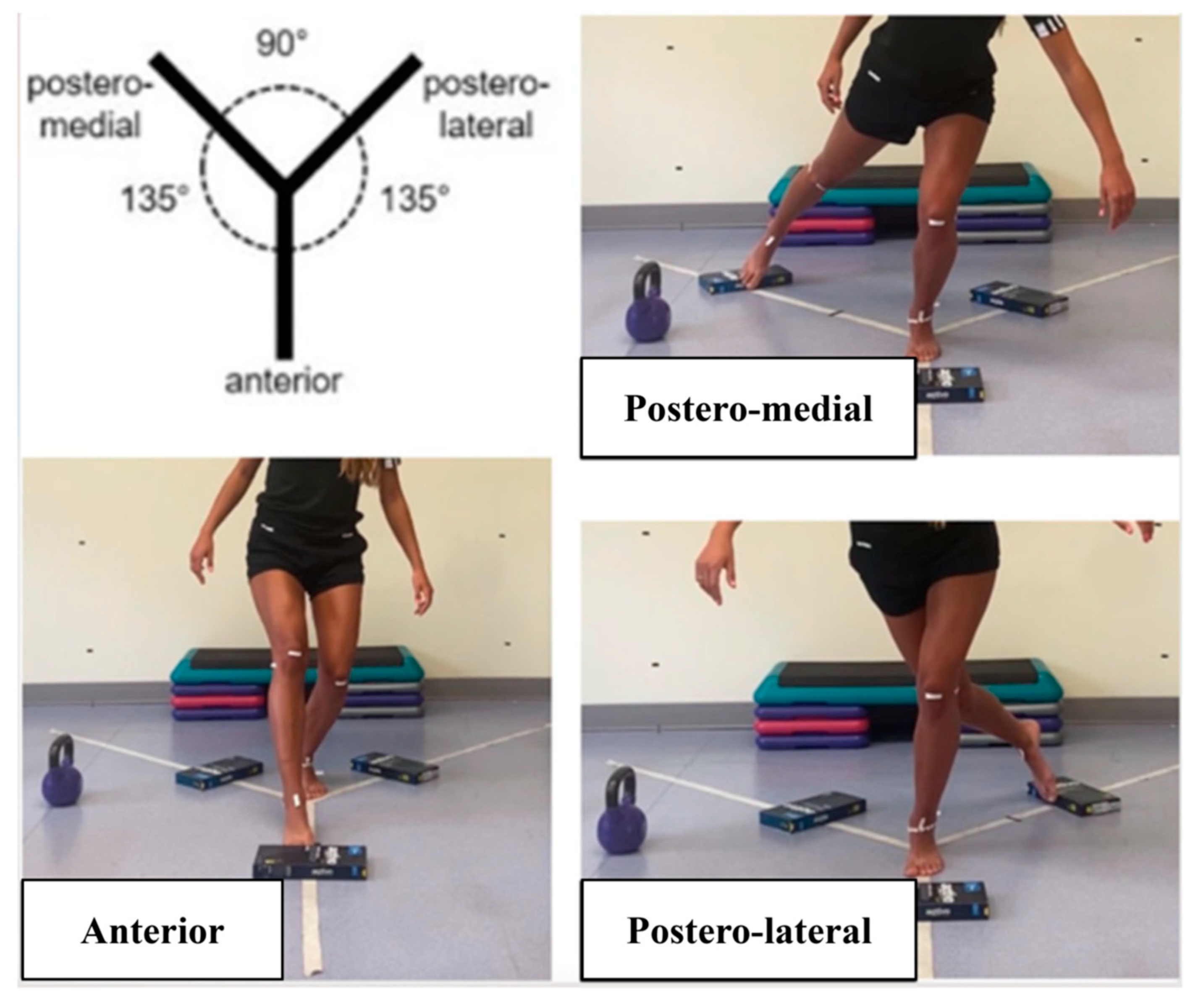
2.5.2. Y Balance Test (YBT)

The YBT evaluates the stability and joint mobility of the ankle, knee, and hip. This test was performed with the participant standing on one leg, placing the support foot in the center of a Y-shaped area formed by three bands on the floor, forming an angle of 135° and 90° between each one. While balancing on one leg, the participant strove to reach as far as possible with the other limb without falling along each of the three branches of the Y: anterior, posterior-right, and posterior-left. Measurements (in cm) were taken from the tip of the foot in each direction, with three trials conducted to obtain an average. The attempt was invalid if the participant supported the free leg during the exercise, lifted, or lost balance. In this study, a kit was used that the athlete had to move, with contact always with the element. (Figure 3) [31].

2.5.3. Lateral Step Down (LSD)

In the LSD test, the participants performed the test unilaterally on a 20 cm step, crossing their arms crossed on the shoulders and standing with the test leg on the edge of the box. The contralateral leg hung off the side of the step with the knee fully extended. Then, they were instructed to lower themselves and bend the test leg until the contralateral leg touched the floor, without losing balance. In this test, knee valgus was evaluated (Figure 4) [32].

2.5.4. Single Leg Squat (SLS)

In the SLS, the participants stood on one leg and were instructed to cross their arms over their chest, flex their nonstanding knee to 90°, and squat down as low as possible without losing balance, with good weight transfer. Then, they had to ascend back to the standing position without contacting the floor with the nontesting leg. The objective of this test was to evaluate knee valgus [33].

2.5.5. Jump Landing Test (JLT)

The JLT was divided into two phases. First, the participant had to perform a horizontal jump forward, and then immediately perform a vertical jump. The starting position was standing on a step, with a mark on the ground at the average height of the participant. Each participant then had to jump longer than the mark for the attempt to be valid. The knee valgus was analyzed in the first standing position (P0), in the first landing position (P1), and in the last landing position (P1) (Figure 5) [34].

2.6. Development of the Assessment Protocol and Intervention

An ultrasound study of the knee was performed to measure the movement of the tibia relative to the femur before and after KT application. Following this measure, the participants performed a previous warm-up of 10 min, including mobility exercises of the joints of the lower limbs, squats, and jump squats. Then, the investigators supervised the functional tests in the following order: YBT, LSD, SLS, and JLT. Before starting the functional tests, the investigators placed markers in the following anatomical areas of the lower limb of each participant: anterior superior iliac spines, knee (central, internal, and external part), malleoli, and astragalus.
Functional tests were performed three times on each leg, first without a bandage and then with the bandage on. Each participant tried each exercise before performing the test to check their adequate execution. The researchers recorded the tests in frontal view to analyze the knee valgus with the ImageJ Software version 2.0 [35]. To systematize data extraction, a 7-step image measurement protocol was designed: (i) calibration of the image from pixels to cm from the patella; (ii) activation of the tool that allows measurement of angles; (iii) delimitation of the longitudinal references lines from the anterior superior iliac spine to the patella, (iv) and from the patella to the astragalus; (v) fitting of the longitudinal reference lines on the anatomical lines; (vi) analysis; (vii) measure. The angle measured was the dynamic knee valgus, which is the angle between the line formed between the anterior superior iliac spine and the middle of the tibiofemoral joint and the line formed from the middle of the tibiofemoral joint to the middle of the ankle mortise [36].
From this moment on, the KT bandage was applied always by the same researcher. In the initial position for the bandage application with the knee flexed at 45°, the KT was applied progressively with the corresponding tension (75% IG; 10% CG) as the participant fully extended the leg. The KT bandage was placed on the anterior tibial tuberosity and moved up cranially. The % tension refers to the % additional length that the KT acquires when stretched (Figure 6) [37,38,39].

2.7. Statistical Analysis

The statistical analysis was performed using the Statistical Package of Social Sciences program (SPSS 25.0v from IBM, Armonk, NY, USA). First, the Shapiro–Wilk test was carried out to check the normal distribution of quantitative variables of the study. Then, a descriptive analysis of the participants was performed using the mean and standard deviation for the parametric variables as well as the median and interquartile range for the non-parametric variables. Thereafter, a two-way analysis of variance (ANOVA) with repeated measures was performed to determine the time x group effects (p-txg) of the intervention. The statistical significance level was set at p < 0.05. Partial eta squared (η2p) was used to measure the effect size, considering 0.01 as small effect size, 0.06 as moderate effect size, and 0.13 as large effect size [40].

3. Results

3.1. Ultrasonographic Variables

Comparing the CG with the IG in the time x group analysis, no statistically significant differences were found in any of the three movements of the tibia: anterior displacement (p-txg = 0.43; η2p = 0.14), posterior displacement (p-txg = 0.43; η2p = 0.04), and total displacement (p-txg = 0.67; η2p = 0.01). In the time analysis, there was a significant difference in the total displacement of the tibia (p = 0.02) (Table 2).

3.2. Functional Tests

During the LSD test, there was a significant difference in the time x group interaction between the IG and CG with a large effect size (p-txg < 0.05, η2p = 0.26) (IG-T1: 151.40 ± 11.04°; IG-T2: 157.10 ± 10.18° versus GC-T1: 156.96 ± 5.44; GC-T2: 158.68 ± 6.12), so the 75% tension KT bandage reduced the valgus of the knee. No statistically significant differences were observed in terms of time x group interaction in the YBT anterior (p-txg = 0.76; η2p = 0.01), YBT medial (p-txg = 0.93; η2p = 0.00), YBT lateral (p-txg = 0.53; η2p = 0.06), SLS (p-txg = 0.17, η2p = 0.10), JLT position 0 (p-txg = 0.51; η2p = 0.03), position 1 (p-txg = 0.20; η2p = 0.09), or position 2 (p-txg = 0.63; η2p = 0.01) (Table 3). In the time analysis, there was a significant difference in the YBT anterior (p = 0.01), the SLS (p < 0.01), and the LSD (p < 0.01).

4. Discussion

This novel pilot study aimed to analyze the immediate effects of a KT bandage on knee valgus, balance, proprioception, and anterior displacement of the tibia in female athletes. The KT application was observed to reduce knee valgus in some functional tests, which could have a preventive effect against ACL injury.
The KT placed with a tension of 75% significantly decreased knee valgus during the LSD test, compared to the KT with 10% tension (p-txg < 0.01), with a large effect size, as opposed to what other studies have reported about the effects of KT on static stability in healthy young athletes [24]. However, in the YBT, significant differences were only found over time in the anterior displacement, but not between groups, which is in line with the results reported by Espí-López et al. in healthy amateur soccer players [24]. Only in studies performed in individuals with a recent ACL rupture did KT improve the static stability [22,23]. This finding is clinically relevant because it may be associated with a potential reduction in the risk of ACL injury, since an enhanced knee valgus is a risk factor for this type of injury [26]. Concerning the SLS, no statistically significant differences were found between bandages, perhaps due to the small sample size (n = 20), as the sample size calculation indicates that it should reach at least n = 50 to obtain significant improvements between groups. Indeed, as there were statistically significant differences (p < 0.01) in the analysis over time, we could assume that, for example, in the SLS test (p-txg = 0.17), statistically significant differences between groups would be found if the sample was larger. However, the trend in the LSD and the SLS is similar, which could be because the muscle pattern of both tests is a closed kinetic chain of lower limbs, with activation of hip extensors, hip abductors, knee extensors, and ankle extensors.
Regarding the JLT, no statistically significant differences between groups were found in the present study. Other authors have reported that KT at the knee reduces the dynamic knee valgus during some landing tasks such as the single leg drop landing test in male participants [25], the Drop Jump test in both female and male participants [20], and Drop Jump test in male participants applying a similar KT bandage to that in the present study [26]. Perhaps the JLT has different biomechanics as it has a horizontal jump component, which could explain why the same results were not found as in other studies.
Concerning the displacement of the tibia, the use of the KT is based on the tactile stimulation of the cutaneous mechanoreceptors, which may improve proprioception and knee position during dynamic tasks by allowing a better perception of the position of the joints. This specific application can help control the anterior translation of the tibia and valgus. In the temporal analysis of the ultrasound assessment, the KT bandage reduced the total displacement of the tibia (p = 0.02). However, there were no statistically significant differences between the IG and the CG, which could be explained by the highly elastic properties of the KT, which allows a normal full range of motion while providing support [19]. In this regard, both KTs with 75% and 10% could be effective.
Some interesting clinical implications can be drawn from this study, which was conducted to find immediate means of prevention for ACL injuries in female athletes following the research recommendations of Limroongreungrat et al. [26]. A 75% tension KT bandage reduced knee valgus during a dynamic stability test in a closed kinetic chain, so it could be an effective strategy to prevent ACL injuries in female rugby and football athletes, combined with other approaches such as proprioception or jumping tasks training [41]. However, it is important to point out the limitations found in the study. First, the sample size is small, which makes it difficult to project the results to the specific population. Second, the observations related only to the direct effects of KT, but the long-term effects are unknown. Considering that this study was performed only with female athletes, it would be essential to consider the hormonal aspect in future research. Current knowledge shows that each person has differences in terms of ligament laxity, but there are intrapersonal differences based on the menstrual cycle [42]. In addition, the knee, as an intermediate joint, depends on the other joints, such as the hip and the foot [43,44,45]. It must be considered that the body is an entity, and in future studies, neighboring joints such as the ankle, hip, or pelvis could be analyzed to carry out a more complete biomechanical study. Finally, in the evaluation of biases, it should be considered that the participants may have noticed the difference in tension between one bandage and another, and these thoughts could interfere with the results.

5. Conclusions

A KT bandage was able to reduce knee valgus compared to a placebo bandage in the LSD test but not in more intense movements and was not more effective at reducing the anterior translation of the tibia. Based on these results, it could have a modest preventive effect against an ACL injury in female athletes. Future experimental designs with larger samples should be carried out with the aim of contracting the effect of the use of the KT bandage in the prevention of valgus collapse of the knee as well as clarifying the effect on restriction of tibiofemoral displacement.

Author Contributions

A.F. and J.M. were in charge of recruitment and assessment of the participants, and prepared the initial draft of the manuscript; J.A.-P. was in charge of assessment and methodology supervision and contributed to the final version of the manuscript; G.G.-P.-d.-S. analyzed and interpreted the data, supervised the methodology, and prepared the final version of the manuscript. All authors have read and agreed to the published version of the manuscript.

Funding

This research received no external funding.

Institutional Review Board Statement

The study was conducted in accordance with the Declaration of Helsinki, and the protocol was approved by the Ethics Committee of the European University of Madrid (CIPI/23.052), 13 February 2023.

Informed Consent Statement

Informed consent was obtained from all subjects involved in the study.

Data Availability Statement

The data presented in this study are available on request from the corresponding author.

Conflicts of Interest

The authors declare that there are no interests that could be perceived as a conflict with respect to the research, authorship, and/or publication of this article.

References

  1. Markatos, K.; Kaseta, M.K.; Lallos, S.N.; Korres, D.S.; Efstathopoulos, N. The anatomy of the ACL and its importance in ACL reconstruction. Eur. J. Orthop. Surg. Traumatol. 2013, 23, 747–752. [Google Scholar] [CrossRef] [PubMed]
  2. Acevedo, R.J.; Rivera-Vega, A.; Miranda, G.; Micheo, W. Anterior Cruciate Ligament Injury: Identification of Risk Factors and Prevention Strategies. Curr. Sports Med. Rep. 2014, 13, 186–191. [Google Scholar] [CrossRef] [PubMed]
  3. Bisciotti, G.N.; Chamari, K.; Cena, E.; Bisciotti, A.; Bisciotti, A.; Corsini, A.; Volpi, P. Anterior cruciate ligament injury risk factors in football. J. Sports Med. Phys. Fit. 2019, 59, 1724–1738. [Google Scholar] [CrossRef] [PubMed]
  4. Griffin, L.Y.; Albohm, M.J.; Arendt, E.A.; Bahr, R.; Beynnon, B.D.; Demaio, M.; Dick, R.W.; Engebretsen, L.; Hannafin, J.A.; Hewett, T.E.; et al. Understanding and preventing noncontact anterior cruciate ligament injuries: A review of the Hunt Valley II meeting, January 2005. Am. J. Sports Med. 2006, 34, 1512–1532. [Google Scholar] [CrossRef] [PubMed]
  5. Bram, J.T.; Magee, L.C.; Mehta, N.N.; Patel, N.M.; Ganley, T.J. Anterior Cruciate Ligament Injury Incidence in Adolescent Athletes: A Systematic Review and Meta-analysis. Am. J. Sports Med. 2021, 49, 1962–1972. [Google Scholar] [CrossRef] [PubMed]
  6. Montalvo, A.M.; Schneider, D.K.; Yut, L.; Webster, K.E.; Beynnon, B.; Kocher, M.S.; Myer, G.D. “What’s my risk of sustaining an ACL injury while playing sports?” A systematic review with meta-analysis. Br. J. Sports Med. 2019, 53, 1003–1012. [Google Scholar] [CrossRef] [PubMed]
  7. Montalvo, A.M.; Schneider, D.K.; Webster, K.E.; Yut, L.; Galloway, M.T.; Heidt, R.S.; Kaeding, C.C.; Kremcheck, T.E.; Magnussen, R.A.; Parikh, S.N.; et al. Anterior Cruciate Ligament Injury Risk in Sport: A Systematic Review and Meta-Analysis of Injury Incidence by Sex and Sport Classification. J. Athl. Train. 2019, 54, 472–482. [Google Scholar] [CrossRef] [PubMed]
  8. Chia, L.; Silva, D.D.O.; Whalan, M.; McKay, M.J.; Sullivan, J.; Fuller, C.W.; Pappas, E. Non-contact Anterior Cruciate Ligament Injury Epidemiology in Team-Ball Sports: A Systematic Review with Meta-analysis by Sex, Age, Sport, Participation Level, and Exposure Type. Sports Med. 2022, 52, 2447–2467. [Google Scholar] [CrossRef]
  9. Waldén, M.; Hägglund, M.; Magnusson, H.; Ekstrand, J. ACL injuries in men’s professional football: A 15-year prospective study on time trends and return-to-play rates reveals only 65% of players still play at the top level 3 years after ACL rupture. Br. J. Sports Med. 2016, 50, 744–750. [Google Scholar] [CrossRef]
  10. Voskanian, N. ACL Injury prevention in female athletes: Review of the literature and practical considerations in implementing an ACL prevention program. Curr. Rev. Musculoskelet. Med. 2013, 6, 158–163. [Google Scholar] [CrossRef]
  11. Hewett, T.E.; Di Stasi, S.L.; Myer, G.D. Current concepts for injury prevention in athletes after anterior cruciate ligament reconstruction. Am. J. Sports Med. 2013, 41, 216–224. [Google Scholar] [CrossRef] [PubMed]
  12. Gobbi, A.; Francisco, R. Factors affecting return to sports after anterior cruciate ligament reconstruction with patellar tendon and hamstring graft: A prospective clinical investigation. Knee Surg. Sports Traumatol. Arthrosc. 2006, 14, 1021–1028. [Google Scholar] [CrossRef] [PubMed]
  13. Silvers-Granelli, H.J.; Bizzini, M.; Arundale, A.; Mandelbaum, B.R.; Snyder-Mackler, L. Does the FIFA 11+ Injury Prevention Program Reduce the Incidence of ACL Injury in Male Soccer Players? Clin. Orthop. Relat. Res. 2017, 475, 2447–2455. [Google Scholar] [CrossRef] [PubMed]
  14. Webster, K.E.; Hewett, T.E. Meta-analysis of meta-analyses of anterior cruciate ligament injury reduction training programs. J. Orthop. Res. 2018, 36, 2696–2708. [Google Scholar] [CrossRef] [PubMed]
  15. Williams, S.; Whatman, C.; Hume, P.A.; Sheerin, K. Kinesio Taping in Treatment and Prevention of Sports Injuries. Sports Med. 2012, 42, 153–164. [Google Scholar] [CrossRef] [PubMed]
  16. Howe, A.; Campbell, A.; Ng, L.; Hall, T.; Hopper, D. Effects of two different knee tape procedures on lower-limb kinematics and kinetics in recreational runners. Scand. J. Med. Sci. Sports 2015, 25, 517–524. [Google Scholar] [CrossRef]
  17. Villar, P.L.; Cabello, M.G.; Medigure, X.M.d.S.P.d.; Marne, P.S.C. Revisión del Kinesio Taping o vendaje neuromuscular como forma de tratamiento fisioterapéutico. Cuest. Fisioter. Rev. Univ. Inf. Investig. Fisioter. 2011, 40, 65–76. [Google Scholar]
  18. de Jesus, J.F.; Novello, A.d.A.; Nakaoka, G.B.; dos Reis, A.C.; Fukuda, T.Y.; Bryk, F.F. Kinesio taping effect on quadriceps strength and lower limb function of healthy individuals: A blinded, controlled, randomized, clinical trial. Phys. Ther. Sport 2016, 18, 27–31. [Google Scholar] [CrossRef]
  19. Lyman, K.J.; Keister, K.; Gange, K.; Mellinger, C.D.; A Hanson, T. Investigating the effectiveness of kinesio® taping space correction method in healthy adults on patellofemoral joint and subcutaneous space. Int. J. Sports Phys. Ther. 2017, 12, 250–257. [Google Scholar]
  20. Rajasekar, S.; Kumar, A.; Patel, J.; Ramprasad, M.; Samuel, A.J. Does Kinesio taping correct exaggerated dynamic knee valgus? A randomized double blinded sham-controlled trial. J. Bodyw. Mov. Ther. 2018, 22, 727–732. [Google Scholar] [CrossRef]
  21. Kielė, D.; Solianik, R. Four-Week Application of Kinesiotaping Improves Proprioception, Strength, and Balance in Individuals With Complete Anterior Cruciate Ligament Rupture. J. Strength Cond. Res. 2023, 37, 213–219. [Google Scholar] [CrossRef] [PubMed]
  22. Bischoff, L.; Babisch, C.; Babisch, J.; Layher, F.; Sander, K.; Matziolis, G.; Pietsch, S.; Röhner, E. Effects on proprioception by Kinesio taping of the knee after anterior cruciate ligament rupture. Eur. J. Orthop. Surg. Traumatol. 2018, 28, 1157–1164. [Google Scholar] [CrossRef] [PubMed]
  23. Ogrodzka-Ciechanowicz, K.; Głąb, G.; Ślusarski, J.; Gądek, A.; Nawara, J. Does kinesiotaping can improve static stability of the knee after anterior cruciate ligament rupture? A randomized single-blind, placebo-controlled trial. BMC Sports Sci. Med. Rehabil. 2021, 13, 24. [Google Scholar] [CrossRef] [PubMed]
  24. Espí-López, G.V.; Serra-Añó, P.; Cobo-Pascual, D.; Zarzoso, M.; Suso-Martí, L.; Cuenca-Martínez, F.; Inglés, M. Effects of Taping and Balance Exercises on Knee and Lower-Extremity Function in Amateur Soccer Players: A Randomized Controlled Trial. J. Sport Rehabil. 2019, 29, 626–632. [Google Scholar] [CrossRef] [PubMed]
  25. Sheikhi, B.; Letafatkar, A.; Hogg, J.; Naseri-Mobaraki, E. The influence of kinesio taping on trunk and lower extremity motions during different landing tasks: Implications for anterior cruciate ligament injury. J. Exp. Orthop. 2021, 8, 25. [Google Scholar] [CrossRef] [PubMed]
  26. Limroongreungrat, W.; Boonkerd, C. Immediate effect of ACL kinesio taping technique on knee joint biomechanics during a drop vertical jump: A randomized crossover controlled trial. BMC Sports Sci. Med. Rehabil. 2019, 11, 32. [Google Scholar] [CrossRef]
  27. Martonick, N.; Kober, K.; Watkins, A.; DiEnno, A.; Perez, C.; Renfro, A.; Chae, S.; Baker, R. The Effect of Kinesio Tape on Factors for Neuromuscular Control of the Lower-Extremity: A Critically Appraised Topic. J. Sport Rehabil. 2020, 29, 841–846. [Google Scholar] [CrossRef] [PubMed]
  28. Haynes, A.B.; Haukoos, J.S.; Dimick, J.B. TREND Reporting Guidelines for Nonrandomized/Quasi-Experimental Study Designs. JAMA Surg. 2021, 156, 879–880. [Google Scholar] [CrossRef]
  29. Lee, P.H.; Macfarlane, D.J.; Lam, T.H.; Stewart, S.M. Validity of the International Physical Activity Questionnaire Short form (IPAQ-SF): A Systematic Review. Int. J. Behav. Nutr. Phys. Act. 2011, 8, 115. [Google Scholar] [CrossRef]
  30. Stoianov, A.G.; Pătrașcu, J.M.; Hogea, B.G.; Andor, B.; Mișcă, L.C.; Florescu, S.; Onofrei, R.R.; Pătrașcu, J.M. Dynamic Ultrasound Assessment of the Anterior Tibial Translation for Anterior Cruciate Ligament Tears Diagnostic. J. Clin. Med. 2022, 11, 2152. [Google Scholar] [CrossRef]
  31. Powden, C.J.; Dodds, T.K.; Gabriel, E.H. The Reliability of the Star Excursion Balance Test and Lower Quarter Y-Balance Test in Healthy Adults: A systematic review. Int. J. Sports Phys. Ther. 2019, 14, 683–694. [Google Scholar] [CrossRef] [PubMed]
  32. Mansfield, C.; Rethman, K.; Stephens, J.; Di Stasi, S.; Vanetten, L.; Briggs, M. The association and reliability of the frontal plane projection angle during the lateral step down test on knee function in patients with patellofemoral pain. Knee 2022, 36, 87–96. [Google Scholar] [CrossRef] [PubMed]
  33. Ugalde, V.; Brockman, C.; Bailowitz, Z.; Pollard, C.D. Single leg squat test and its relationship to dynamic knee valgus and injury risk screening. PM&R 2015, 7, 229–235. [Google Scholar] [CrossRef]
  34. Mehl, J.; Diermeier, T.; Herbst, E.; Imhoff, A.B.; Stoffels, T.; Zantop, T.; Petersen, W.; Achtnich, A. Evidence-based concepts for prevention of knee and ACL injuries. 2017 guidelines of the ligament committee of the German Knee Society (DKG). Arch. Orthop. Trauma Surg. 2018, 138, 51–61. [Google Scholar] [CrossRef]
  35. Schroeder, A.B.; Dobson, E.T.A.; Rueden, C.T.; Tomancak, P.; Jug, F.; Eliceiri, K.W. The ImageJ ecosystem: Open-source software for image visualization, processing, and analysis. Protein Sci. 2021, 30, 234–249. [Google Scholar] [CrossRef]
  36. Munro, A.; Herrington, L.; Carolan, M. Reliability of 2-dimensional video assessment of frontal-plane dynamic knee valgus during common athletic screening tasks. J. Sport Rehabil. 2012, 21, 7–11. [Google Scholar] [CrossRef]
  37. Ogrodzka-Ciechanowicz, K.; Stolarz, M.; Gła̧b, G.; Ślusarski, J.; Ga̧dek, A. Biomechanical image of the knee motion in patients with chronic anterior instability of the knee joint before and after Kinesio Taping. J. Back Musculoskelet. Rehabil. 2020, 33, 169–177. [Google Scholar] [CrossRef]
  38. Kase, K.; Wallis, J.; Kase, T. Clinical Therapeutic Applications of the Kinesio Taping Method, 2nd ed.; Albuquerque, Kinesio Taping Assoc: Tokyo, Japan, 2003. [Google Scholar]
  39. Almazán-Polo, J.; López-López, D.; Romero-Morales, C.; Rodríguez-Sanz, D.; Becerro-De-Bengoa-Vallejo, R.; Losa-Iglesias, M.E.; Bravo-Aguilar, M.; Calvo-Lobo, C. Quantitative Ultrasound Imaging Differences in Multifidus and Thoracolumbar Fasciae between Athletes with and without Chronic Lumbopelvic Pain: A Case-Control Study. J. Clin. Med. 2020, 9, 2647. [Google Scholar] [CrossRef] [PubMed]
  40. Lakens, D. Calculating and reporting effect sizes to facilitate cumulative science: A practical primer for t-tests and ANOVAs. Front. Psychol. 2013, 4, 62627. [Google Scholar] [CrossRef]
  41. Johnson, J.L.; Capin, J.J.; Arundale, A.J.; Zarzycki, R.; Smith, A.H.; Snyder-Mackler, L. A Secondary Injury Prevention Program May Decrease Contralateral Anterior Cruciate Ligament Injuries in Female Athletes: 2-Year Injury Rates in the ACL-SPORTS Randomized Controlled Trial. J. Orthop. Sports Phys. Ther. 2020, 50, 523–530. [Google Scholar] [CrossRef]
  42. Moriceau, J.; Fevre, A.; Domínguez-Balmaseda, D.; González-De-La-Flor, Á.; Simón-Areces, J.; García-Pérez-De-Sevilla, G. The Influence of the Menstrual Cycle and Oral Contraceptives on Knee Laxity or Anterior Cruciate Ligament Injury Risk: A Systematic Review. Appl. Sci. 2022, 12, 12627. [Google Scholar] [CrossRef]
  43. Boden, B.P.; Sheehan, F.T.; Torg, J.S.; Hewett, T.E. Noncontact anterior cruciate ligament injuries: Mechanisms and risk factors. J. Am. Acad. Orthop. Surg. 2010, 18, 520–527. [Google Scholar] [CrossRef] [PubMed]
  44. Sahabuddin, F.N.A.; Jamaludin, N.I.; Amir, N.H.; Shaharudin, S. The effects of hip- and ankle-focused exercise intervention on dynamic knee valgus: A systematic review. PeerJ 2021, 9, e11731. [Google Scholar] [CrossRef] [PubMed]
  45. Hewett, T.E.; Myer, G.D. The mechanistic connection between the trunk, hip, knee, and anterior cruciate ligament injury. Exerc. Sport Sci. Rev. 2011, 39, 161–166. [Google Scholar] [CrossRef] [PubMed]
Figure 1. Position of the participant during the measurement with ultrasonography.
Applsci 14 02839 g001
Figure 2. Measurements of the anteroposterior displacement of the tibia. (A) The initial position of the previous displacement. (B) Anterior displacement of the tibia with respect to the femur. (C) The initial position of posterior displacement. (D) Posterior displacement of the tibia with respect to the femur.
Applsci 14 02839 g002
Figure 3. Y Balance Test.
Applsci 14 02839 g003
Figure 4. Lateral Step Down Test.
Applsci 14 02839 g004
Figure 5. Jump Landing Test.
Applsci 14 02839 g005
Figure 6. KT bandage application.
Applsci 14 02839 g006
Table 1. Demographic characteristics of the sample (n = 10).
VariablesMin–MaxMean (SD)
Age (years)18–3121.90 ± 3.48
Height (cm)159–171165.00 ± 0.04
Weight (kg)50–6857.55 ± 5.63
BMI (kg/m2)18.56–25.9121.10 ± 2.55
Physical activity level (MET/min/week)1653–46922861.10 ± 949.62
SD, standard deviation; BMI, body mass index; MET, metabolic equivalent.
Table 2. Ultrasonographic variables for comparisons of tibiofemoral displacement between groups.
VariablesGROUPT1T2p-Timep-txgη2p txg
Anterior displacement (cm)IG
CG
0.13 ± 0.11
0.10 ± 0.09
0.08 ± 0.04
0.08 ± 0.05
0.100.430.14
Posterior displacement (cm)IG
CG
0.08 ± 0.07
0.10 ± 0.05
0.05 ± 0.03
0.06 ± 0.05
0.100.430.04
Total displacement (cm)IG
CG
0.21 ± 0.17
0.20 ± 0.13
0.13 ± 0.05
0.15 ± 0.05
0.02 *0.670.01
T1, initial assessment; T2, final assessment; IG, intervention group (75% tension KT); CG, control group 10% tension KT); p-time: p-value time; p-txg: p-value time × group assessment. η2p txg: effect size for the time × group interaction. All the analyses were performed with a two-way ANOVA. Significance was set at p < 0.05 (bold) *.
Table 3. Comparisons between groups in the knee valgus angle and performance during functional tests.
VariablesGROUPT1T2p-Timep-txgη2p txg
Y balance TestANT (cm)IG
CG
61.95 ± 4.73
62.47 ± 4.55
60.03 ± 4.90
63.01 ± 3.74
0.01 *0.760.01
MED (cm)IG
CG
71.93 ± 6.31
73.38 ± 5.71
73.96 ± 6.91
75.21 ± 7.49
0.130.930.00
LAT (cm)IG
CG
76.15 ± 8.58
76.22 ± 8.19
76.82 ± 6.09
78.76 ± 6.73
0.290.530.06
LSD (degrees)IG
CG
151.40 ± 11.04
156.96 ± 5.44
157.10 ± 10.18
158.68 ± 6.12
<0.01 *0.02 *0.26
SLS (degrees)IG
CG
167.55 ± 12.03
169.27 ± 6.50
173.32 ± 10.50
172.75 ± 5.86
<0.01 *0.170.10
JLTP0 (degrees)IG
CG
178.87 ± 7.40
180.10 ± 9.01
178.83 ± 6.29
181.90 ± 6.90
0.530.510.03
P1 (degrees)IG
CG
186.66 ± 10.96
185.95 ± 13.85
184.42 ± 12.59
188.50 ± 9.16
0.930.200.09
P2 (degrees)IG
CG
185.68 ± 6.26
183.73± 4.57
185.77 ± 8.76
184.95 ± 7.70
0.570.630.01
T1, initial assessment; T2, final assessment; IG, intervention group (75% tension KT); CG, control group (10% tension KT); ANT, anterior; MED, medial; LAT, lateral; LSD, lateral step down; SLS, single leg squat; JLT, jump landing test; P0, starting position; P1, landing 1; P2, landing 2; p-time: p-value time; p-txg: p-value time × group assessment. η2p txg: effect size for the time x group interaction. All the analysis were performed with a two-way ANOVA. Significance was set at p < 0.05 (* bold).
Disclaimer/Publisher’s Note: The statements, opinions and data contained in all publications are solely those of the individual author(s) and contributor(s) and not of MDPI and/or the editor(s). MDPI and/or the editor(s) disclaim responsibility for any injury to people or property resulting from any ideas, methods, instructions or products referred to in the content.

