Fanconi – Bickel Syndrome – two cases report - edigraphic.com
Fanconi – Bickel Syndrome – two cases report - edigraphic.com
Fanconi – Bickel Syndrome – two cases report - edigraphic.com

You also want an ePaper? Increase the reach of your titles
YUMPU automatically turns print PDFs into web optimized ePapers that Google loves.
Annals<br />
of<br />
Hepatology<br />
Abstract<br />
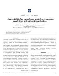
Annals N of Sotelo Hepatology et al. <strong>Fanconi</strong> 2008; 7(2): – <strong>Bickel</strong> April-June: <strong>Syndrome</strong>163-167 163<br />
Case Report<br />
<strong>Fanconi</strong> – <strong>Bickel</strong> <strong>Syndrome</strong> – <strong>two</strong> <strong>cases</strong> <strong>report</strong><br />
Norberto Sotelo; 1 Ramiro García; 2 René Tostado; 3 Nagasharmila Dhanakotti 4<br />
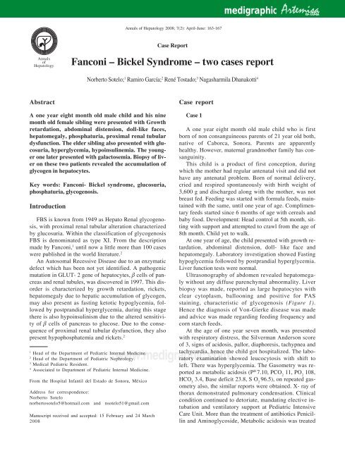
A one year eight month old male child and his nine<br />
month old female sibling were presented with Growth<br />
retardation, abdominal distension, doll-like faces,<br />
hepatomegaly, phosphaturia, proximal renal tubular<br />
dysfunction. The elder sibling also presented with glucosuria,<br />
hyperglycemia, hypoinsulinemia. The younger<br />
one later presented with galactosemia. Biopsy of liver<br />
on these <strong>two</strong> patients revealed the accumulation of<br />
glycogen in hepatocytes.<br />
Key words: <strong>Fanconi</strong>- <strong>Bickel</strong> syndrome, glucosuria,<br />
phosphaturia, glycogenosis.<br />
Introduction<br />
FBS is known from 1949 as Hepato Renal glycogenosis,<br />
with proximal renal tubular alteration characterized<br />
by glucosuria. Within the classification of glycogenosis<br />
FBS is denominated as type XI. From the description<br />
made by <strong>Fanconi</strong>, 1 until now a little more than 100 <strong>cases</strong><br />
were published in the world literature. 2<br />
An Autosomal Recessive Disease due to an enzymatic<br />
defect which has been not yet identified. A pathogenic<br />
mutation in GLUT- 2 gene of hepatocytes, β cells of pancreas<br />
and renal tubules, was discovered in 1997. This disorder<br />
is characterized by growth retardation, rickets,<br />
hepatomegaly due to hepatic accumulation of glycogen,<br />
may also present as fasting ketotic hypoglycemia, followed<br />
by postprandial hyperglycemia, during this stage<br />
there is also hypoinsulinism due to the altered sensitivity<br />
of β cells of pancreas to glucose. Due to the consequence<br />
of proximal renal tubular dysfunction, they also<br />
present hypophosphatemia and rickets. 2<br />
1 Head of the Department of Pediatric Internal Medicine.<br />
2 Head of the Department of Pediatric Nephrology.<br />
3 Medical Pediatric Resident.<br />
4 Associated to Department of Pediatric Internal Medicine.<br />
From the Hospital Infantil del Estado de Sonora, México<br />
Address for correspondence:<br />
Norberto Sotelo<br />
norbertosotelo5@hotmail.<strong>com</strong> and nsotelo51@gmail.<strong>com</strong><br />
Manuscript received and accepted: 15 February and 24 March<br />
2008<br />
Case <strong>report</strong><br />
Case 1<br />
www.m<strong>edigraphic</strong>.<strong>com</strong><br />
Artemisa<br />
m<strong>edigraphic</strong> en línea<br />
A one year eight month old male child who is first<br />
born of non consanguineous parents of 21 year old both,<br />
native of Caborca, Sonora. Parents are apparently<br />
healthy. However, maternal grandmother family has consanguinity.<br />
This child is a product of first conception, during<br />
which the mother had regular antenatal visit and did not<br />
have any antenatal problem. Born of normal delivery,<br />
cried and respired spontaneously with birth weight of<br />
3,600 g and discharged along with the mother, was not<br />
breast fed. Feeding was started with formula feeds, maintained<br />
with the same, until one year of age. Complimentary<br />
feeds started since 6 months of age with cereals and<br />
baby food. Development: Head control at 5th month, sitting<br />
with support and attempted to crawl from the age of<br />
8th month. Child yet to walk.<br />
At one year of age, the child presented with growth retardation,<br />
abdominal distension, doll- like face and<br />
hepatomegaly. Laboratory investigation showed Fasting<br />
hypoglycemia followed by postprandial hyperglycemia.<br />
Liver function tests were normal.<br />
Ultrasonography of abdomen revealed hepatomegaly<br />
without any diffuse parenchymal abnormality. Liver<br />
biopsy was made, <strong>report</strong>ed as large hepatocytes with<br />
clear cytoplasm, ballooning and positive for PAS<br />
staining, characteristic of glycogenosis (Figure 1).<br />
Hence the diagnosis of Von-Gierke disease was made<br />
and advice was made regarding feeding frequency and<br />
corn starch feeds.<br />
At the age of one year seven month, was presented<br />
with respiratory distress, the Silverman Anderson score<br />
of 3, signs of acidosis, pallor, diaphoresis, tachypnea and<br />
tachycardia, hence the child got hospitalized. The labo-<br />
ratory examination showed leucocytosis with shift to<br />
left. There was hyperglycemia. The Gasometry was <strong>report</strong>ed<br />
as metabolic acidosis (P H 7.10, PCO 2 11, PO 2 108,<br />
HCO 3 3.4, Base deficit 23.8, S O 2 96.5), on repeated gasometry<br />
also, the similar <strong>report</strong>s were obtained. X- ray of<br />
thorax demonstrated pulmonary condensation. Clinical<br />
condition continued to detoriate, mandating elective intubation<br />
and ventilatory support at Pediatric Intensive<br />
Care Unit. More than the treatment of antibiotics Penicillin<br />
and Aminoglycoside, Metabolic acidosis was treated
164<br />
Case 1<br />
Case 2<br />
with continuous bicarbonate infusion calculated as per<br />
acid base deficit depending upon the gasometry. Due to<br />
persistent hyperglycemia, determination of C peptide<br />
level was requested, with result of 0.3 ng/mL, with reference<br />
value of 1.1- 5.0 ng/mL; Insulin level < 0.200μU/L<br />
with reference value of 5.0- 25μU/L. The antibiotic<br />
scheme was modified, started on Dicloxacillin, Cefotaxim<br />
and Fluconazole as Urine culture was positive for<br />
Candida Albicans of 100,000/HPF. With this, the child<br />
improved clinically, radiologically and values of laboratory<br />
examination, hence extubated and transferred to us<br />
for continued evaluation and treatment.<br />
The physical examination showed, Head circumfer-<br />
ence of 48 cm ( 50th centile for age), length of 73 c (<<br />
5th centile for age), weight of 11,100 g (< 25th centile<br />
for age); Doll-like face was more prominent, small thorax<br />
and rachitic changes noted in the costal cartilage. Distended<br />
abdomen with hepatomegaly of 6 x 5 x 5 cm<br />
(Figure 2), with genitals relevant to his age and sex also<br />
noted. Rest of the systemic examination was normal.<br />
Repeat Gasometry, C peptide, Insulin were requested<br />
and were continued to be below the normal values<br />
(metabolic acidosis, 0.3 ng/mL and 1.3μU/L respec-<br />
Annals of Hepatology 7(2) 2008: 163-167<br />
A B<br />
A B<br />
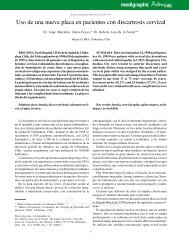
Figure 1. Liver tissue of Case 1 and Case 2: (A) PAS staining demonstrating glycogen accumulation and (B) Photograph of positive diastase<br />
test.<br />
www.m<strong>edigraphic</strong>.<strong>com</strong><br />
tively). Hence, the possibility of FBS associated with<br />
glycogenosis was considered. Due to persistent acidosis,<br />
Nephrology and Genetic opinion was sought.<br />
Serum and urinary electrolytes of 24 hours were repeated.<br />
While analyzing Phosphaturia, Negative anion<br />
gap curve of Hyperchloremic type was observed,<br />
this was <strong>com</strong>patible with tubulopathies (Figure 3<br />
and Table I).<br />
Radiography of long bones showed evidence of rickets.<br />
Ultrasonography of Abdomen showed hepatomegaly<br />
without diffuse hepatic parenchymal lesions. Kidneys,<br />
Pancreas and Spleen were normal in shape and size.<br />
In the beginning, the diagnosis of type I glycogenosis<br />
Von Gierke Disease was considered in this patient. How-<br />
ever, during the course of evolution, the patient presented<br />
with metabolic acidosis, due to renal tubular dysfunction,<br />
pre-prandial hypoglycemia with ketosis followed<br />
by postprandial elevated blood glucose , associated decreased<br />
Insulin and C Peptide levels, but at the same time<br />
not following the clinical course of type I diabetes too.<br />
After the biochemical analysis and renal functional test<br />
to demonstrate the proximal renal tubular dysfunction,<br />
with the results of glucosuria, phosphaturia, bicarbonate
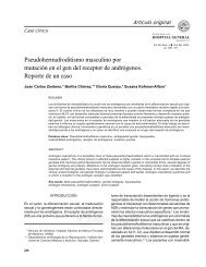
Figure 2. Case 1 showing typical Doll-like face and hepatomegaly<br />
(marked by lines).<br />
Table I. Laboratory test.<br />
loss, hypoglycemia, hypophosphatemia, hypocalcemia,<br />
hypoglycemia with radiological evidence of rickets, the<br />
diagnosis of FBS was made in this case as regard to the<br />
clinical picture.<br />
The treatment was started with 25 hydroxy Vitamin<br />
D (0.25μg/24 h or 2,000 MI/kg/24), Disodium phosphate<br />
with Phosphoric acid as 85% (58.5 g in 1,200<br />
mL of normal water) 5 times a day and Bicarbonate of<br />
15 mEq/k/24 h. Dietary advice was made as mater-<br />
N Sotelo et al. <strong>Fanconi</strong> – <strong>Bickel</strong> <strong>Syndrome</strong> 165<br />
Anion Gap<br />
www.m<strong>edigraphic</strong>.<strong>com</strong><br />
50<br />
40<br />
30<br />
20<br />
10<br />
0<br />
2<br />
0 10 20 30 40 50 60<br />
Proportion of urinary chloride<br />
Figure 3. Demonstrating the displacement of anion gap curve<br />
towards left (hyperchloremic) in proportion to urinary chloride<br />
(1- Case 1; 2 – Case 2). AG = urinary Na + urinary K – urinary Cl<br />
(Case 1: 87 + 14 = 101-57 = +44; Case 2: 32 + 23 = 52-23 =<br />
+32).<br />
Blood/Serum Urine<br />
Reference<br />
Parameter Case 1 Case 2 Reference value Parameter Case 1 Case 2 value<br />
Arterial blood P H 7.10 7.33 7.35 – 7.45 Sodium (mEq/L) 87 32 85 – 250<br />
Sodium (mEq/L) 142 131 135 – 145 Potasium (mEq/L) 14 23 2.5 - 125<br />
Potassium (mEq/L) 3.9 3.9 3.5 – 5.0 Chloride (mEq/L) 57 23 15 - 40<br />
Chloride (mEq/L) 110 98 95 – 105 Phosphorous (mg/dL) 28.4 23 0.6 – 1.5<br />
Bicarbonate (mEq/L) 9 9.3 22 – 26 Glucose (mg/dL) 1,000 Negative Negative<br />
Calcium (mg/dL) 9 8.9 8.8 – 9.5 Ketone(Mm/dL) 150 Negative or Neg<br />
Phosphorous (mg/dL) 2.3 2.2 4.0 – 7.0 Amino acid Negative Negative Negative<br />
Urea (mg/dL) 11 14 15 – 40 Chromatography<br />
Creatinine (mg/dL) 0.4 0.1 0.6 – 1.2<br />
Glucose (mg/dL) 117 117 70 - 110<br />
Uric acid (mg/dL) 1.5 – 2.0 – 2.7<br />
Alkaline phosphatase IU 14 12 30 – 120<br />
Cholesterol (mg/dL) 125 139 < 200<br />
Trigliceride (mg/dL) 370 215 85 – 150<br />
Insulin (uU/L) 0.20 1.9 5.0 – 25<br />
C Peptide (ng/mL) 0.3 0.41 1.1 – 5.0<br />
Alanin amino tranferase U/L 51.0 37.0 0-38.0<br />
Asparte amino transferase U/L 37.0 28.0 7- 41<br />
Prothrombin time (sec) 14 .0 13.0 11 – 14<br />
Thrombo plastin time (sec) 24 .0 21.0 20 – 40<br />
Total Bilirubin (mg/dL) 0.7 1.4 0-1.1<br />
Direct Bilirubin (mg/dL) 0.1 0.7 0-0.3<br />
Unconjugated Bilirubin (mg/dL) 0.6 0.7 0-0.9<br />
Total protein (g/dL) 7.6 6.8 6-8.7<br />
Galactose (mg/dL) 4.5 46.8 0-5.0<br />
nized cow’s milk in the beginning, later whole milk<br />
was given as the galactose level in the blood was normal.<br />
In addition to that, fruits, frequent small feeds<br />
and use of corn starch in the diet of the child also re<strong>com</strong>mended.<br />
The diet was calculated to provide the calorie<br />
of 140 kcal/kg of body weight and 5 grams of protein/kg<br />
of body weight. Regular follow up on outpatient<br />
basis was advised with Pediatric and Nephrology<br />
departments.<br />
1
166<br />
Case 2<br />
Eight month old female infant, who is born of second<br />
conception (sibling of case 1), delivered via naturalis<br />
with birth weight of 2,500 g, length of 48 cm and head<br />
circumference of 32 cm. During the neonatal period, was<br />
hospitalized for transient tachypnea, transient unconjugated<br />
hyperbilirubinemia and hypoglycemia not associated<br />
with seizures. The newborn metabolic screening was<br />
negative, however the estimation of blood galactose level<br />
later <strong>report</strong>ed to be elevated.<br />
At the age of 7 month, infant was brought to hospital<br />
for delay attaining age appropriate milestones (not attained<br />
head control, not started sitting and crawling).<br />
The physical examination revealed weight of 7,950<br />
g, length of 63 cm, which are less for age (3 rd centile),<br />
Doll-like face, prominent abdomen and hepatomegaly<br />
Figure 4. Case 2 exhibits Doll-like face and hepatomegaly (marked<br />
by lines).<br />
Case 1<br />
Annals of Hepatology 7(2) 2008: 163-167<br />
www.m<strong>edigraphic</strong>.<strong>com</strong><br />
of about 6 cm below the right costal margin (Figure 4).<br />
Because of these finding, FBS was suspected and the<br />
following laboratory analysis were requested: Arterial<br />
blood gas, Serum and Urinary Electrolytes, calcium,<br />
phosphorous, cholesterol, triglyceride, ultra sonogram<br />
of abdomen and x-ray of thorax and long bone. They<br />
were <strong>report</strong>ed as metabolic acidosis (Table I), rickets<br />
and hepatomegaly devoid of any specific ultrasonographic<br />
pattern. Based on these results, liver biopsy was<br />
done and revealed histopathological changes typical of<br />
glycogenosis (Figure 1). Hence, the suspicion of FBS<br />
was confirmed and treatment was continued with phosphate<br />
and bicarbonate replacement. The dietary advice<br />
was given based on the calculation of 140 kcal/kg of<br />
body weight, protein of 4.5 gm/kg of body weight in<br />
the form of maternized cow’s milk, formula of soya, puree<br />
of fruits and vegetables.<br />
Discussion<br />
FBS is a rare clinical entity and rare presentation with<br />
in the same family affecting the infants between the ages<br />
of 2 month to 1 year of life. The clinical picture, which is<br />
noticed in majority of the patients, is characteristic faces<br />
«Moon- shaped» face or «Doll- like» face. In addition,<br />
short stature, rickets, protuberant abdomen, and<br />
hepatomegaly 3-5 are associated features in FBS. In addition,<br />
biochemical changes encountered as a consequence<br />
of the altered Proximal Renal Tubular function (glucosuria,<br />
phosphaturia, bicarbonate loss and hypophosphatemia,<br />
aminoaciduria). Preprandial ketotic hypoglycemia<br />
and significant post prandial hyperglycemia and<br />
hypergalactesemia are noted due to low hepatic uptake.<br />
There is also hypoinsulinemia due to altered sensitivity<br />
of β cells of Pancreas to glucose, associated with reduced<br />
release of monosachroide by hepatic cells during fasting<br />
Case 2<br />
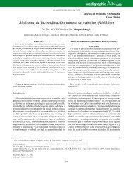
Figure 5. Percutaneous liver biopsy of Case 1 and Case 2 (HE 40x) showed large and ballooned hepatocytes with clear cytoplasm.
and intra cellular accumulation with inhibition of glycogenolysis<br />
leading on to glycogen accumulation and<br />
hepatomegaly.<br />
ESTE DOCUMENTO ES ELABORADO POR MEDI-<br />
GRAPHIC<br />
6,7<br />
During the period of fasting, there is reduced blood<br />
glucose level which is generally not associated with convulsions.<br />
This hypoglycemia is ac<strong>com</strong>panied by ketosis<br />
followed by postprandial hyperglycemia, making to consider<br />
the possibility of ketosis to have a neuro protective<br />
effect. 2,8 It is also possible to encounter elevation of cholesterol<br />
and triglyceride in these patients due to accumulation<br />
of secondary metabolite. 6,7<br />
With respect to recent discovery in Genetic aspect of<br />
the disease, mutations in GLUT-2 gene was detected in<br />
34 <strong>cases</strong>. 7,9,10 Parental consanguinity was observed in 2/<br />
3rd of these patients. Some of these <strong>cases</strong> were identified<br />
through Neonatal Screening for Galactosemia. This gene<br />
is localized much in hepatocytes like in the β cells of<br />
pancreas, enterocytes and renal tubules. 2<br />
There is no specific treatment exist for this disease<br />
in the therapeutic form, besides intravenous hydrations,<br />
to provide with Vitamin D in the form of 1, 25<br />
dihydroxy Vitamin D3. Citrate up to 15 mEq/kg/day<br />
given every 4th hourly to maintain bicarbonate more<br />
than 20 mEq/dL, phosphate solution orally; to offer<br />
with fructose and corn starch to provide glucose in<br />
slow release form and also which do not require diseased<br />
metabolic pathway. Because of the possible association<br />
of Galactosemia with FBS, it is re<strong>com</strong>mend-<br />
N Sotelo et al. <strong>Fanconi</strong> – <strong>Bickel</strong> <strong>Syndrome</strong> 167<br />
www.m<strong>edigraphic</strong>.<strong>com</strong><br />
ed to provide the diet free of Galactose, until the results<br />
of metabolic studies are known. 2,6,10<br />
References<br />
1. <strong>Fanconi</strong> G, <strong>Bickel</strong> H. Die chroische Aminoacidurie<br />
(Aminosaeurediabetes oder nephrotischglukourischer<br />
Zwergwuchs) bei der Glykogenoseund der Cystinkrankheit. Helv<br />
Pediat Acta 1949; 4: 359-396.<br />
2. Pascual JM. Hereditary syndrome of glucose transport. Med Clin<br />
(Barc.) 2006; 127(18): 709-714.<br />
3. Garty R, Cooper M, Taibachnik E. The <strong>Fanconi</strong> syndrome associated<br />
with hepatic glycogenosis and abnormal metabolism of<br />
galactose. J Pediatr 1974; 85: 821-823.<br />
4. Velasquez JL. Síndrome de <strong>Fanconi</strong>. Bol Med Hosp Infant Mex<br />
1987; 44: 509-510.<br />
5. Santer R, Schneppenheim R, Suter D, Schanb J, Steinman B.<br />
<strong>Fanconi</strong>-<strong>Bickel</strong> syndrome- the original patient and his natural<br />
history, historical steps leading to the primary defect, and a review<br />
of the literature. Eur J Pediatr 1998; 157: 783-797.<br />
6. Velasquez-Jones L, Portillo HV, Sanjines R, Gamboa-Marrufo<br />
A, Feria-Kaiser K, Valencia-Mayoral P. Síndrome de <strong>Fanconi</strong>-<br />
<strong>Bickel</strong>. Bol Med Hosp Infant Mex 1991; 48: 255-259.<br />
7. Santer R, Steinmann B, Schaub J. <strong>Fanconi</strong>-<strong>Bickel</strong> syndrome a<br />
congenital defect of facilitative glucose transport. Curr Mol Med<br />
2002; 2: 213-227.<br />
8. De Vivo D, Lackie M, Ferrendelli J, McDougal D. Chronic ketosis<br />
and cerebral metabolism. Ann Neurol 1987; 3: 331-337.<br />
9. Sakamoto O, Ogawa E, Ohura T, Igarashi Y, Matsubara Y,<br />
Narisawa K, et al. Mutation analysis of the GLUT 2 gene in<br />
patients with fanconi-<strong>Bickel</strong> syndrome. Pediatr Res 2000; 48:<br />
586-589.<br />
10. Karande S, Kumbhare N, Kulkarni M. <strong>Fanconi</strong>-<strong>Bickel</strong> syndrome.<br />
Indian Pediatrics 2007; 44: 223-225.