Heberprot-P: A Novel Product for Treating Advanced Diabetic Foot ...
Heberprot-P: A Novel Product for Treating Advanced Diabetic Foot ...
Heberprot-P: A Novel Product for Treating Advanced Diabetic Foot ...

Create successful ePaper yourself
Turn your PDF publications into a flip-book with our unique Google optimized e-Paper software.
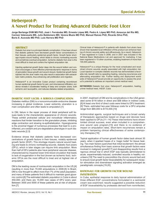
<strong>Heberprot</strong>-P:<br />
A <strong>Novel</strong> <strong>Product</strong> <strong>for</strong> <strong>Treating</strong> <strong>Advanced</strong> <strong>Diabetic</strong> <strong>Foot</strong> Ulcer<br />
DIABETIC FOOT ULCER: A SERIOUS COMPLICATION<br />
Diabetes mellitus (DM) is a noncommunicable endocrine disease<br />
increasing in global incidence. Lower extremity ulceration is a<br />
main complication and often leads to amputation.[1]<br />
In DM, failure in the repair process of distal peripheral soft tissues<br />
leads to the characteristic appearance of chronic wounds.<br />
These exhibit protracted cellular and noncellular infl ammatory<br />
reactions that hinder transition to the granulation phase, inhibiting<br />
edge contraction and slowing re-epithelialization. Hyperglycemia<br />
is the proximal trigger of numerous processes that lead to a proinfl<br />
amed, pro-oxidant and pro-degradative phenotype in such diabetic<br />
wounds.[2,3]<br />
Evidence shows that diabetic patients have decreased concentrations<br />
of growth factors in their tissues, notably epidermal<br />
growth factor (EGF). This shortage impairs natural wound healing<br />
and leads to chronic nonhealing wounds, diabetic foot ulcers<br />
(DFU), which in later stages can require limb amputation. More<br />
than half of DFU patients also have peripheral vascular disease,<br />
characterized by impaired lower limb blood circulation that leads<br />
to lack of oxygenation in the foot (known as ischemic foot). Ischemic<br />
DFUs are the most diffi cult to treat and at highest risk of<br />
amputation.<br />
DM is the leading cause of nontraumatic amputation in the US,[4]<br />
resulting in more than 70,000 amputations in 2008.[5] In Brazil,<br />
DM is now thought to affect more than 7% of the adult population,<br />
and many of these patients fi nd it diffi cult to maintain good glycemic<br />
control.[6] The estimated diabetic population in Cuba is about<br />
450,000; and there are 15,000 new cases of DFU every year.<br />
Between 3000 and 5000 of these patients are at risk of amputation.[7]<br />
MEDICC Review, January 2013, Vol 15, No 1<br />
Special Article<br />
Jorge Berlanga DVM MS PhD, José I. Fernández MD, Ernesto López MS, Pedro A. López MD PhD, Amaurys del Río MD,<br />
Carmen Valenzuela MS, Julio Baldomero MD, Verena Muzio MD PhD, Manuel Raíces PhD, Ricardo Silva PhD,<br />
Boris E. Acevedo MD PhD, Luis Herrera MD PhD<br />
ABSTRACT<br />
<strong>Diabetic</strong> foot ulcer is a principal diabetic complication. It has been shown<br />
that diabetic patients have decreased growth factor concentrations in<br />
their tissues, particularly epidermal growth factor. Growth factor shortage<br />
impairs wound healing, which leads to chronic nonhealing wounds<br />
and sometimes eventual amputation. Ischemic diabetic foot ulcer is the<br />
most diffi cult to treat and confers the highest amputation risk.<br />
Injecting epidermal growth factor deep into the wound bottom and contours<br />
encourages a more effective pharmacodynamic response in terms<br />
of granulation tissue growth and wound closure. Epidermal growth factor<br />
injected into the ulcer matrix may also result in association with extracellular<br />
matrix proteins, thus enhancing cell proliferation and migration.<br />
<strong>Heberprot</strong>-P is an innovative Cuban product containing recombinant<br />
human epidermal growth factor <strong>for</strong> peri- and intra-lesional infi ltration; evidence<br />
reveals it accelerates healing of deep and complex ulcers, both<br />
ischemic and neuropathic, and reduces diabetes-related amputations.<br />
Clinical trials of <strong>Heberprot</strong>-P in patients with diabetic foot ulcers have<br />
shown that repeated local infi ltration of this product can enhance healing<br />
of chronic wounds safely and effi caciously. As a result, <strong>Heberprot</strong>-<br />
P was registered in Cuba in 2006, and in 2007 was included in the<br />
National Basic Medications List and approved <strong>for</strong> marketing. It has<br />
been registered in 15 other countries, enabling treatment of more than<br />
100,000 patients.<br />
<strong>Heberprot</strong>-P is a unique therapy <strong>for</strong> the most complicated and recalcitrant<br />
chronic wounds usually associated with high amputation risk.<br />
Local injection in complex diabetic wounds has demonstrated a favorable<br />
risk–benefi t ratio by speeding healing, reducing recurrences and<br />
attenuating amputation risk. Further testing and deployment worldwide<br />
of <strong>Heberprot</strong>-P would provide an opportunity to assess the product’s<br />
potential to address an important unmet medical need.<br />
KEYWORDS <strong>Diabetic</strong> foot ulcer, <strong>Heberprot</strong>-P, amputation, healing,<br />
unmet medical need, rhEGF, Cuba<br />
In 2007, treatment of DM and its complications in the USA generated<br />
some $116 billion in direct and $58 billion in indirect costs.<br />
[8] At least one third of direct costs were linked to DFU treatment.<br />
[9] There, estimated two-year followup costs <strong>for</strong> a DFU amputee<br />
range from $80,000 to $110,000.[10]<br />
Antimicrobial agents, surgical techniques and a broad variety<br />
of therapeutic approaches based on drugs and devices have<br />
been applied to DFUs.[11–14] These interventions have shown<br />
limited clinical success, even when included in a comprehensive<br />
wound care program,[15] and there is no evidence of<br />
impact on amputation rates. Short-term recurrences remain a<br />
problem hampering clinical effectiveness of some contemporary<br />
therapies.[16]<br />
Topical application of human growth factor dates back almost 30<br />
years, when it sparked hopes of a ‘magic bullet’ <strong>for</strong> tissue healing.<br />
Two main factors quenched that initial excitement: the almost<br />
simultaneous fi nding from basic science that growth factors were<br />
involved in malignant growth[17] and disappointing results from a<br />
rigorous clinical trial in which EGF was topically administered to<br />
acute, experimentally-induced, controlled wounds in healthy volunteers.[18]<br />
The need to precondition the chronic wound bed and<br />
to ensure local growth factor bioavailability <strong>for</strong> subsequent receptor<br />
stimulation and downstream signaling activation emerged as<br />
paradigmatic concepts.[19,20]<br />
RATIONALE FOR GROWTH FACTOR WOUND<br />
INFILTRATION<br />
In Cuba, epithelial response to daily topical administration of<br />
three different EGF concentrations <strong>for</strong>mulated in a semisolid<br />
cream was examined; results suggested a possible reduction<br />
of EGF bioavailability by proteases derived from noninfected,<br />
Peer Reviewed<br />
11
Special Article<br />
acute, controlled wounds.[21] This was somewhat surprising<br />
as other studies had already established proteolysis affecting<br />
growth factors and their receptors in chronic circumstances.[22,23]<br />
It is worth noting that previous studies had<br />
also documented the need <strong>for</strong> prolonged interaction between<br />
EGF and its receptor to achieve a significant granulation tissue<br />
response in controlled wounds in mice.[24] Our initial<br />
research indicated 125 I-EGF was rapidly cleared from the<br />
application site, probably by protease-driven cleavage and<br />
receptor-mediated endocytosis. Mean residence time values<br />
suggested that over 60% of the amount administered could<br />
have disappeared as early as two hours after administration.<br />
[25] The message of these studies was that even acute, clean<br />
and controlled wounds may not represent a hospitable substrate<br />
<strong>for</strong> growth factor physical and chemical integrity. Previous<br />
disappointing clinical results may have been due to local<br />
bioavailability limitations.[26,27]<br />
Such knowledge prompted the hypothesis that injecting EGF<br />
deep into the wound base and walls would allow <strong>for</strong> greater<br />
pharmacodynamic response in terms of granulation tissue<br />
growth and wound closure. In further studies, single or repeated<br />
EGF systemic or local injections produced clear-cut cytoprotective<br />
and proliferative responses, suggesting an intrinsic ability<br />
of EGF at supraphysiological concentrations to trigger biological<br />
events necessary <strong>for</strong> tissue repair.[28–30]<br />
Injecting EGF into the tissue, down and inside the base and<br />
walls (including the dermo-epidermal junction), possibly also<br />
reduces its degradation following topical application and<br />
contact with wound exudate. These experiments identified<br />
three layers of cellular response potential along the longitudinal<br />
axis of granulation tissue. Fibroblasts populating<br />
the more superficial stratum expressed far more prohibitin<br />
and far less EGF receptor. <strong>Advanced</strong> glycosilated endproducts<br />
and elastase also appeared overexpressed next to the<br />
wound surface than in deeper cells strata. It is likely that<br />
topographic positioning along the wound bed axis dictates<br />
fibroblasts’ intrinsic ability to respond to a mitogenic signal.<br />
Notably, prohibitin is a renowned inhibitor of cell cycle progression.[31]<br />
Contemporary evidence supports that EGF<br />
injected into the ulcer matrix may result in an association<br />
complex with extracellular matrix proteins, thus enhancing<br />
cell proliferation and migration.[32]<br />
Classic studies have shown that growth factor effectively<br />
counteracts senescence of chronic ulcer-derived fi broblasts—<br />
including diabetic ulcer fi broblasts—and stimulates proliferation.[33,34]<br />
Appropriate wound bed preparation through sharp<br />
debridement and infection elimination is required prior to infi ltration.<br />
HEBERPROT-P IMPROVES HEALING AND REDUCES<br />
AMPUTATIONS IN PATIENTS WITH SEVERE DFU<br />
Following earlier research, scientists at the Center <strong>for</strong> Genetic<br />
Engineering and Biotechnology (CIGB, the Spanish acronym) in<br />
Havana developed <strong>Heberprot</strong>-P, a patented pharmaceutical composition<br />
whose parenteral <strong>for</strong>mulation is based on rhEGF. The<br />
product is administered in DFU patients by intralesional infi ltration<br />
to accelerate healing of deep and complex ulcers, either neuropathic<br />
or ischemic.[35]<br />
12<br />
Peer Reviewed<br />
Based on the rationale that rhEGF can enhance healing of chronic<br />
wounds following repeated local infi ltrations,[36] various clinical<br />
trials using <strong>Heberprot</strong>-P in DFU patients have been conducted,<br />
demostrating safety and effi cacy.[37–43] Infi ltration with rhEGF<br />
<strong>for</strong> diabetic wound healing does not replace standard procedures<br />
but should be incorporated into comprehensive wound care along<br />
with medical interventions to correct patients’ glycemia and creatinine.<br />
In a compassionate study with terminal ulcer patients in 2001–<br />
2002, the fi rst clinical evidence using EGF infi ltration <strong>for</strong> diabetic<br />
foot ulcers and amputation residual bases emerged.[37] All lesions<br />
were chronic, complex and recalcitrant, Wagner scale stages 3<br />
and 4.[36] Effi cacy demonstrated in these types of wounds paved<br />
the way <strong>for</strong> solid clinical development, which culminated in a<br />
nationwide, double-blind, placebo-controlled phase III clinical trial,<br />
duly registered with the appropriate Cuban regulatory agency.[39]<br />
Since then, EGF local injection has been used <strong>for</strong> complex diabetic<br />
wounds in various Cuban clinical trials, demonstrating a<br />
favorable risk–benefi t balance by speeding healing, reducing<br />
recurrences and attenuating amputation risk.[43] Adverse effects<br />
were preponderantly mild to moderate (65.6% mild, 28.6% moderate,<br />
and only 3.7% severe), with pain and burning sensation<br />
at administration site the most frequent. Pain reported was mild<br />
to moderate in intensity and was not associated with treatment<br />
suspension. A dose-effect relation associated with appearance of<br />
shivering and chills was consistently obtained in all trials at both<br />
doses used (25 μg and 75 μg) and in the pooled analysis; intensity<br />
was mild to moderate and symptom appearance was not associated<br />
with treatment suspension.[39]<br />
EGF infi ltration increased and accelerated healing in poor-prognosis<br />
wounds toward a rapid and sustained response (Figure 1).<br />
More than 80% granulation was obtained globally with <strong>Heberprot</strong>-<br />
P, in comparison with less than 60% with standard care alone. Of<br />
patients treated with <strong>Heberprot</strong>-P at 75 μg, three times per week<br />
until complete granulation (or during 8 weeks) in association with<br />
standard care, 77% healed; while only 56% healed with placebo<br />
injections and standard care.[40] Seminal clinical trials are summarized<br />
in Table 1.<br />
As a result, <strong>Heberprot</strong>-P was registered in Cuba in 2006, and<br />
in 2007 was included in the National Basic Medications List<br />
and approved <strong>for</strong> marketing. <strong>Heberprot</strong>-P has also been registered<br />
in 15 other countries (Table 2) enabling treatment of over<br />
100,000 patients. Registration and market approval submissions<br />
are in process in countries such as Brazil, Russia, China,<br />
South Africa, and the Arab states of the Persian Gulf. A Spanish<br />
phase II clinical trial <strong>for</strong> the DFU indication, approved by the<br />
Spanish Drug Agency under European Good Clinical Practices,<br />
concluded recently (publication pending), with the aim of moving<br />
to a pivotal phase III clinical trial in Europe.<br />
EFFECTIVE COMPREHENSIVE DFU TREATMENT:<br />
AN UNMET MEDICAL NEED<br />
Adjuvant therapies and advanced technologies can be used in<br />
addition to standard care as a second line of treatment when<br />
appropriate. These include some topical drugs but are mostly<br />
medical devices: living skin equivalents, specialized dressings,<br />
hyperbaric oxygen therapy and negative pressure devices. These<br />
interventions provide moderate improvement over standard treat-<br />
MEDICC Review, January 2013, Vol 15, No 1
Figure 1: Severe diabetic foot ulcers treated with <strong>Heberprot</strong>-P<br />
Ischemic Patient A<br />
Be<strong>for</strong>e fi rst injection of <strong>Heberprot</strong>-P: 11.9 cm 2<br />
After seven weeks’ treatment: 1.1 cm 2<br />
At week 11: healed<br />
Photos: Dr A del Río Martín<br />
(available in color online at www.medicc.org/mediccreview/berlanga.html)<br />
Table 1: Seminal clinical trials with <strong>Heberprot</strong>-P<br />
MEDICC Review, January 2013, Vol 15, No 1<br />
Ischemic Patient B<br />
Be<strong>for</strong>e fi rst injection of <strong>Heberprot</strong>-P: 21.8 cm 2<br />
After seven weeks’ treatment: 0.5 cm 2<br />
At week 11: healed<br />
Special Article<br />
Trial Details Results Ref.<br />
Phase I: Exploratory<br />
Phase II: Treatment<br />
dose determination<br />
Phase II: Treatment<br />
dose determination<br />
Phase III: Confi rmatory<br />
trial<br />
Phase IV:<br />
Pharmacovigilance<br />
DFU: diabetic foot ulcer<br />
Patients with poor prognosis, 25 μg three times a week,<br />
until granulation or 8 weeks<br />
25 μg or 75 μg three times a week, until granulation or<br />
8 weeks<br />
25 μg or 75 μg three times a week, until healing or 8<br />
weeks<br />
Double-blind placebo-controlled multicenter study, 25<br />
μg versus 75 μg versus placebo (all 3 times a week)<br />
Good safety pattern<br />
Promising results in granulation, healing and<br />
amputation avoidance<br />
Good safety pattern in both doses<br />
Trend to greater effi cacy at 75 μg<br />
Trend to greater effi cacy when treatment is<br />
prolonged until healing<br />
Confi rm effi cacy (granulation and healing) and<br />
safety in patients with Wagner 3−4 DFUs<br />
Pharmacovigilance study in 1835 patients Confi rmation of safety profi le Unpublished<br />
Peer Reviewed<br />
37<br />
38<br />
39<br />
40<br />
13
Special Article<br />
ments, generally only 15% to 20% healing in less than 20 weeks,<br />
and may be expensive and time consuming.[44] In ischemic<br />
patients, surgical revascularization is not suitable <strong>for</strong> all cases and<br />
Table 2: <strong>Heberprot</strong>-P registration year by country<br />
Country Registration Year<br />
Cuba 2006<br />
Algeria 2008<br />
Argentina 2009<br />
Uruguay 2009<br />
Dominican Republic 2009<br />
Venezuela 2010<br />
Ecuador 2010<br />
Mexico 2010<br />
Paraguay 2010<br />
Libya 2010<br />
Colombia 2011<br />
Guatemala 2011<br />
Georgia 2011<br />
Ukraine 2011<br />
Vietnam 2012<br />
Philippines 2012<br />
REFERENCES<br />
1. Davis WA, Norman PE, Bruce DG, Davis TM.<br />
Predictors, consequences and cost of diabetesrelated<br />
lower extremity amputation complicating<br />
type 2 diabetes: the Fremantle Diabetes Study.<br />
Diabetologia. 2006 Nov;49(11):2634–41.<br />
2. Berlanga J, Schultz G, Lopez P. Biology of the<br />
diabetic wound. In: Overhaussen PE, editor. <strong>Foot</strong><br />
ulcers: causes, diagnosis and treatments. New<br />
York: Nova Science Publishers, Inc.; 2009.<br />
3. Schultz G, Berlanga J, Cowan L, Stechmiller J.<br />
Linking the advanced glycation endproducts/<br />
receptor <strong>for</strong> advanced glycation endproduts pathway<br />
in diabetics with infl ammation and topical<br />
antiinfl ammatory treatments of chronic wound.<br />
In: Chandan S, editor. Advances in Wound Care.<br />
Columbus (US): Ohio State University Medical<br />
Center; 2009. Chapter 44. Vol 1.<br />
4. CDC Online Press Release [Internet]. Atlanta:<br />
Center <strong>for</strong> Disease Control and Prevention (US);<br />
[reviewed 2012 Nov 12; cited 2012 Sep 18].<br />
Available from: www.cdc.gov/diabetes/<br />
5. Li Y, Burrows NR, Gregg EW, Albright A, Geiss<br />
LS. Declining rates of hospitalization <strong>for</strong> nontraumatic<br />
lower-extremity amputation in the diabetic<br />
population aged 40 years or older: U.S., 1988–<br />
2008. Diabetes Care. 2012 Feb;35(2):273–7.<br />
6. International Working Group on the <strong>Diabetic</strong><br />
<strong>Foot</strong> [Internet]. Brussels: International<br />
Diabetes Federation; c2000–2005. <strong>Foot</strong><br />
Note Brazil; [cited 2012 Nov 5]; [about 1<br />
screen]. Available from: http://www.iwgdf.org/<br />
index.php?Itemid=37&id=10&option=com<br />
_content&task=view<br />
7. Anuario Estadístico de Salud 2010. Havana: Ministry<br />
of Public Health (CU); 2011 Apr. Spanish.<br />
8. American Diabetes Association. Economic costs<br />
of diabetes in the U.S. in 2007. Diabetes Care.<br />
2008 Mar;31(3):596–615.<br />
9. Driver VR, Fabbi M, Lavery LA, Gibbons G. The<br />
costs of diabetic foot: the economic case <strong>for</strong> the<br />
limb salvage team. J Am Podiatr Med Assoc.<br />
2010 Sep–Oct;100(5):335–41.<br />
14<br />
10. Carls GS, Gibson TB, Driver VR, Wrobel JS,<br />
Garoufalis MG, Defrancis RR, et al. The economic<br />
value of specialized lower-extremity medical<br />
care by podiatric physicians in the treatment<br />
of diabetic foot ulcers. J Am Podiatr Med Assoc.<br />
2011 Mar–Apr;101(2):93–115.<br />
11. Wieman TJ, Smiell JM, Su Y. Effi cacy and safety<br />
of a topical gel <strong>for</strong>mulation of recombinant human<br />
platelet-derived growth factor-BB (becaplermin)<br />
in patients with chronic neuropathic diabetic<br />
ulcers. A phase III randomized placebo-controlled<br />
double-blind study. Diabetes Care. 1998<br />
May;21(5):822–7.<br />
12. Armstrong DG, Lipsky BA. <strong>Diabetic</strong> foot infections:<br />
stepwise medical and surgical management.<br />
Int Wound J. 2004 Jun;1(2):123–32.<br />
13. Dalla PL, Faglia E. Treatment of diabetic foot<br />
ulcer: an overview strategies <strong>for</strong> clinical approach.<br />
Curr Diabetes Rev. 2006 Nov;2(4):431–47.<br />
14. Ha Van G. [Management of a diabetic foot ulcer].<br />
Rev Med Interne. 2008 Sep;29 Suppl 2:S238–<br />
S42. French.<br />
15. Smiell JM, Wieman TJ, Steed DL, Perry BH,<br />
Sampson AR, Schwab BH. Effi cacy and safety of<br />
becaplermin (recombinant human platelet-derived<br />
growth factor-BB) in patients with nonhealing, lower<br />
extremity diabetic ulcers: a combined analysis<br />
of four randomized studies. Wound Repair Regen.<br />
1999 Sep–Oct;7(5):335–46.<br />
16. Gregor S, Maegele M, Sauerland S, Krahn JF,<br />
Peinemann F, Lange S. Negative pressure<br />
wound therapy: a vacuum of evidence? Arch<br />
Surg. 2008 Feb;143(2):189–96.<br />
17. Berlanga-Acosta J, Gavilondo J, García del<br />
Barco D, Martín J, Guillén G. Epidermal Growth<br />
Factor (EGF) and Platelet-Derived Growth Factor<br />
(PDGF) as tissue healing agents: Clarifying concerns<br />
about their possible role in malignant trans<strong>for</strong>mation<br />
and tumor progression. J Carcinogene<br />
Mutagene. 2011 Feb 25;2(1):100–15.<br />
18. Cohen IK, Crossland MC, Garrett A, Diegelmann<br />
RF. Topical application of epidermal growth factor<br />
Peer Reviewed<br />
some authors consider its effectiveness to be overestimated:[45]<br />
in any case, the regulatory process <strong>for</strong> surgical procedures does<br />
not require the same degree of clinical detail as demanded <strong>for</strong><br />
biological products. There remains a clear need <strong>for</strong> adequate<br />
comprehensive therapy to improve healing in severe wounds, <strong>for</strong><br />
which <strong>Heberprot</strong>-P has demonstrated clear benefi cial potential.<br />
In the USA, 8.3% of the population—25.8 million people—have<br />
DM,[46] and there<strong>for</strong>e an estimated 25% lifetime risk of developing<br />
a DFU.[47] The International Diabetes Federation predicts<br />
that the number of people with DM in the USA will be 36 million in<br />
2030, 12% population prevalence.[48]<br />
The estimated number of US DFU patients in 2010 was between<br />
3.9 and 4.6 million. Among these, 2.5 million patients had concomitant<br />
ischemia and hence were at greater risk of complications.[49]<br />
<strong>Heberprot</strong>-P would address the therapeutic needs of this population,<br />
as well as those at risk globally, especially patients with ischemic<br />
wounds that are the most diffi cult to heal.<br />
Further testing of <strong>Heberprot</strong>-P—a unique and fi rst-in-class<br />
therapy to treat the most complicated and recalcitrant chronic<br />
wounds with a high risk of amputation— would provide an<br />
opportunity to assess the product’s potential to address this<br />
vast unmet medical need in different populations and settings<br />
worldwide.<br />
onto partial-thickness wounds in human volunteers<br />
does not enhance reepithelialization. Plast<br />
Reconstr Surg. 1995 Aug;96(2):251–4.<br />
19. Schultz GS, Sibbald RG, Falanga V, Ayello EA,<br />
Dowsett C, Harding K, et al. Wound bed preparation:<br />
a systematic approach to wound management.<br />
Wound Repair Regen. 2003 Mar;11 Suppl 1:S1–28.<br />
20. Sibbald RG, Orsted H, Schultz GS, Coutts P,<br />
Keast D; International Wound Bed Preparation<br />
Advisory Board. Preparing the wound bed 2003:<br />
focus on infection and infl ammation. Ostomy<br />
Wound Manage. 2003 Nov;49(11):23–51.<br />
21. Berlanga J, Lodos J, Reyes O, Caballero E,<br />
Lopez Saura P. Epidermal growth factor stimulated<br />
re-epithelialization in pigs. The possible role<br />
of acute wound proteases. Biotecnol Aplicada.<br />
1998;15(2):83–7.<br />
22. Saarialho-Kere UK. Patterns of matrix metalloproteinase<br />
and TIMP expression in chronic<br />
ulcers. Arch Dermatol Res. 1998 Jul;290<br />
Suppl:S47–54.<br />
23. Mast BA, Schultz GS. Interactions of cytokines,<br />
growth factors, and proteases in acute and<br />
chronic wounds. Wound Repair Regen. 1996<br />
Oct;4(4):411–20.<br />
24. Buckley A, Davidson JM, Kamerath CD, Wolt TB,<br />
Woodward SC. Sustained release of epidermal<br />
growth factor accelerates wound repair. Proc Natl<br />
Acad Sci USA. 1985 Nov;82(21):7340–4.<br />
25. Prats PA, Duconge J, Valenzuela C, Berlanga<br />
J, Edrosa CR, Fernández-Sánchez E. Disposition<br />
and receptor-site binding of (125)I-EGF after<br />
topical administration to skin wounds. Biopharm<br />
Drug Dispos. 2002 Mar;23(2):67–76.<br />
26. Falanga V, Eaglstein WH, Bucalo B, Katz<br />
MH, Harris B, Carson P. Topical use of human<br />
recombinant epidermal growth factor (h-EGF)<br />
in venous ulcers. J Dermatol Surg Oncol. 1992<br />
Jul;18(7):604–6.<br />
27. Falanga V. Growth factors and chronic wounds:<br />
the need to understand the microenvironment. J<br />
Dermatol. 1992 Nov;19(11):667–72.<br />
MEDICC Review, January 2013, Vol 15, No 1
28. Berlanga-Acosta J, Gavilondo-Cowley J, López-<br />
Saura P, González-López T, Castro-Santana<br />
MD, López-Mola E, et al. Epidermal growth factor<br />
in clinical practice - a review of its biological<br />
actions, clinical indications and safety implications.<br />
Int Wound J. 2009 Oct;6(5):331–46.<br />
29. Berlanga J, Caballero E, Prats P, López Saura<br />
P, Play<strong>for</strong>d RJ. [The role of the epidermal growth<br />
factor in cell and tissue protection]. Med Clin<br />
(Barc). 1999 Sep 4;113(6):222–9. Spanish.<br />
30. Berlanga-Acosta J, Play<strong>for</strong>d RJ, Mandir N,<br />
Goodlad RA. Gastrointestinal cell proliferation<br />
and crypt fi ssion are separate but complementary<br />
means of increasing tissue mass following<br />
infusion of epidermal growth factor in rats. Gut.<br />
2001 Jun;48(6):803–7.<br />
31. Mishra S, Murphy LC, Nyomba BL, Murphy LJ.<br />
Prohibitin: a potential target <strong>for</strong> new therapeutics.<br />
Trends Mol Med. 2005 Apr;11(4):192–7.<br />
32. Hollier B, Harkin DG, Leavesley D, Upton Z.<br />
Responses of keratinocytes to substrate-bound<br />
vitronectin: growth factor complexes. Exp Cell<br />
Res. 2005 Apr 15;305(1):221–32.<br />
33. Stanley AC, Park HY, Phillips TJ, Russakovsky<br />
V, Menzoian JO. Reduced growth of dermal<br />
fi broblasts from chronic venous ulcers can be<br />
stimulated with growth factors. J Vasc Surg. 1997<br />
Dec;26(6):994–9.<br />
34. Loots MA, Lamme EN, Mekkes JR, Bos JD,<br />
Middelkoop E. Cultured fi broblasts from chronic<br />
diabetic wounds on the lower extremity (noninsulin-dependent<br />
diabetes mellitus) show disturbed<br />
proliferation. Arch Dermatol Res. 1999<br />
Feb–Mar;291(2–3):93–9.<br />
35. Berlanga J, Fernández JI, Valdés C, Franco N,<br />
Rojas I, Santana H, et al, inventors; Centro de<br />
Ingeniería Genética Biotechnología, assignee.<br />
Use of a pharmaceutical composition containing<br />
epidermal growth factor (EGF) <strong>for</strong> diabetic foot<br />
amputation prevention. World patent WO PCT/<br />
CU2002/000011. 2008 Dec 18.<br />
36. Berlanga-Acosta J. <strong>Diabetic</strong> lower extremity<br />
wounds: the rationale <strong>for</strong> growth factorsbased<br />
infi ltration treatment. Int Wound J. 2011<br />
Dec;8(6):612–20.<br />
37. Acosta JB, Savigne W, Valdez C, Franco N, Alba<br />
JS, del Río A, et al. Epidermal growth factor<br />
intralesional infi ltrations can prevent amputation<br />
in patients with advanced diabetic foot wounds.<br />
Int Wound J. 2006 Sep;3(3):232–9.<br />
38. Fernández-Montequín JI, Infante-Cristiá E,<br />
Valenzuela-Silva C, Franco-Pérez N, Savigne-<br />
Gutierrez W, Artaza-Sanz H, et al. Intralesional<br />
injections of Citoprot P® (recombinant human<br />
epidermal growth factor) in advanced diabetic<br />
foot ulcers with risk of amputation. Int Wound J.<br />
2007 Dec;4(4):333–43.<br />
MEDICC Review, January 2013, Vol 15, No 1<br />
39. Fernández-Montequín JI, Betancourt BY, Leyva-<br />
Gonzalez G, López Mola E, Galán-Naranjo K,<br />
Ramírez-Navas M, et al. Intralesional administration<br />
of epidermal growth factor-based <strong>for</strong>mulation<br />
(<strong>Heberprot</strong>-P) in advanced diabetic foot ulcer:<br />
Treatment up to complete wound closure. Int<br />
Wound J. 2009 Feb;6(1):67–72.<br />
40. Fernández-Montequín JI, Valenzuela-Silva CM,<br />
González-Díaz O, Savigne W, Sancho-Soutelo<br />
N, Rivero-Fernández F, et al. Intra-lesional<br />
injections of recombinant human Epidermal<br />
growth factor promote granulation and healing in<br />
advanced diabetic foot ulcers. Multicenter, randomized,<br />
placebo-controlled, double blind study.<br />
Int Wound J. 2009 Dec;6(6):432–43.<br />
41. Hernández Rivero MJ, Llanes Barrios JA, Acosta<br />
Lapera DS. [<strong>Heberprot</strong>-P, an effective therapy<br />
<strong>for</strong> prevention of diabetic foot amputation]. Rev<br />
Cubana Angiol Cirugía Vascular. 2009;10(1):3–<br />
11. Spanish.<br />
42. Valenzuela-Silva CM, Tuero-Iglesias AD, García-<br />
Iglesias E, González-Díaz O, del Río-Martín A,<br />
Yera-Alos IB, et al. Granulation Response and<br />
Partial Wound Closure predict Healing in Clinical<br />
Trials on <strong>Advanced</strong> Diabetes <strong>Foot</strong> Ulcers<br />
Treated with Recombinant, Human Epidermal<br />
Growth Factor. Diabetes Care. 2012 Sep 10.<br />
[Epub ahead of print]<br />
43. Ló pez-Saura PA, Berlanga-Acosta J, Ferná ndez-<br />
Montequí n JI, Valenzuela-Silva C, Gonzá lez-Dí az<br />
O, Savigne W, et al. Intralesional Human Recombinant<br />
Epidermal Growth Factor <strong>for</strong> the Treatment<br />
of <strong>Advanced</strong> <strong>Diabetic</strong> <strong>Foot</strong> Ulcer: From Proof<br />
of Concept to Confi rmation of the Effi cacy and<br />
Safety of the Procedure. In: Dinh T, editor. Global<br />
Perspective on <strong>Diabetic</strong> <strong>Foot</strong> Ulcerations. Rijeka<br />
(HR): InTech; 2011 Dec 9. 278 p.<br />
44. Blume P, Driver VR, Tallis AJ, Kirsner RS, Kroeker<br />
R, Payne WG, et al. Formulated collagen<br />
gel accelerates healing rate immediately after<br />
application in patients with diabetic neuropathic<br />
foot ulcers. Wound Repair Regen. 2011 May–<br />
Jun;19(3):302–8.<br />
45. Taylor SM, Johnson BL, Samies NL, Rawlinson<br />
RD, Williamson LE, Davis SA, et al. Contemporary<br />
management of diabetic neuropathic foot<br />
ulceration: a study of 917 consecutively treated<br />
limbs. J Am Coll Surg. 2011 Apr;212(4):532–45.<br />
46. CDC [Internet]. Atlanta (US): Center <strong>for</strong> Disease<br />
Control and Prevention (US); 2012. 2011 National<br />
Diabetes Fact Sheet; [updated 2012 Jan; cited<br />
2012 Sep]; [2.7 MB]. Available from: www.cdc<br />
.gov/diabetes/pubs/pdf/ndfs_2011.pdf<br />
47. Singh N, Armstrong DG, Lipsky BA. Preventing<br />
foot ulcers in patients with diabetes. JAMA. 2005<br />
Jan 12;293(2):217–28.<br />
48. International Diabetes Federation. IDF Diabetes<br />
Atlas, 4th ed. Brussels (BE): International Diabetes<br />
Federation; 2007.<br />
Peer Reviewed<br />
Special Article<br />
49. Yost ML. <strong>Diabetic</strong> <strong>Foot</strong> Ulcers, peripheral artery<br />
disease and critical limb ischemia, 2010. Atlanta,<br />
Georgia: The SAGE Group; 2010.<br />
THE AUTHORS<br />
Jorge Berlanga Acosta, veterinarian with a<br />
master’s degree in pathology and a doctorate in<br />
pharmacology, Center <strong>for</strong> Genetic Engineering<br />
and Biotechnology (CIGB), Havana, Cuba.<br />
José I. Fernández Montequín, angiologist and<br />
vascular surgeon, National Institute <strong>for</strong> Angiology<br />
and Vascular Surgery, Havana, Cuba.<br />
Ernesto López Mola, nutritional pharmacist<br />
with a master’s degree in biotechnology, CIGB,<br />
Havana, Cuba.<br />
Pedro A. López Saura, clinical biochemist with<br />
a doctorate in biology, CIGB, Havana, Cuba.<br />
Amaurys del Río Martín, medical immunologist,<br />
CIGB, Havana, Cuba.<br />
Carmen Valenzuela Silva, mathematician, CIGB.<br />
Julio Baldomero Hernández, family physician,<br />
CIGB, Havana, Cuba.<br />
Verena Muzio González, immunologist with a<br />
doctorate in biology, CIGB, Havana, Cuba.<br />
Manuel Raíces Pérez, biologist. Business and<br />
Projects Development Division, CIGB, Havana,<br />
Cuba.<br />
Ricardo Silva Rodríguez, biologist, CIGB,<br />
Havana, Cuba.<br />
Boris E. Acevedo-Castro (Corresponding<br />
author: boris.acevedo@cigb.edu.cu), physician<br />
with a doctorate in medical sciences, CIGB,<br />
Havana, Cuba.<br />
Luis Herrera Martínez, geneticist with a doctorate<br />
in biology. Director General, CIGB, Havana,<br />
Cuba.<br />
Submitted: September 14, 2012<br />
Approved <strong>for</strong> publication: January 15, 2013<br />
Disclosures: All authors except Fernández are<br />
employed at CIGB, developer of <strong>Heberprot</strong>-P<br />
15